AnatOMEE Project (2023-2025)
Media: Figma, Autodesk Maya, Adobe Illustrator, Adobe After Effects
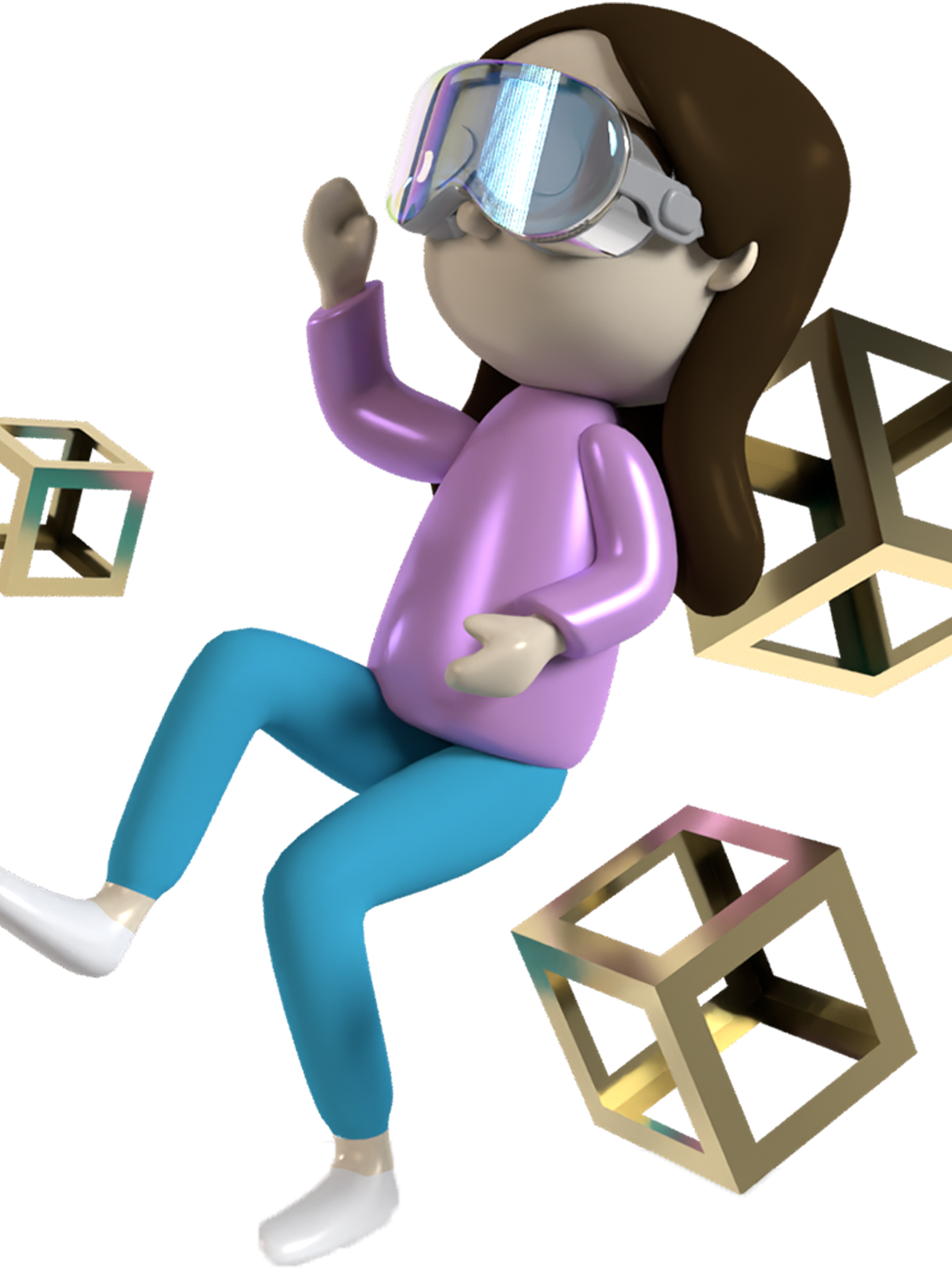
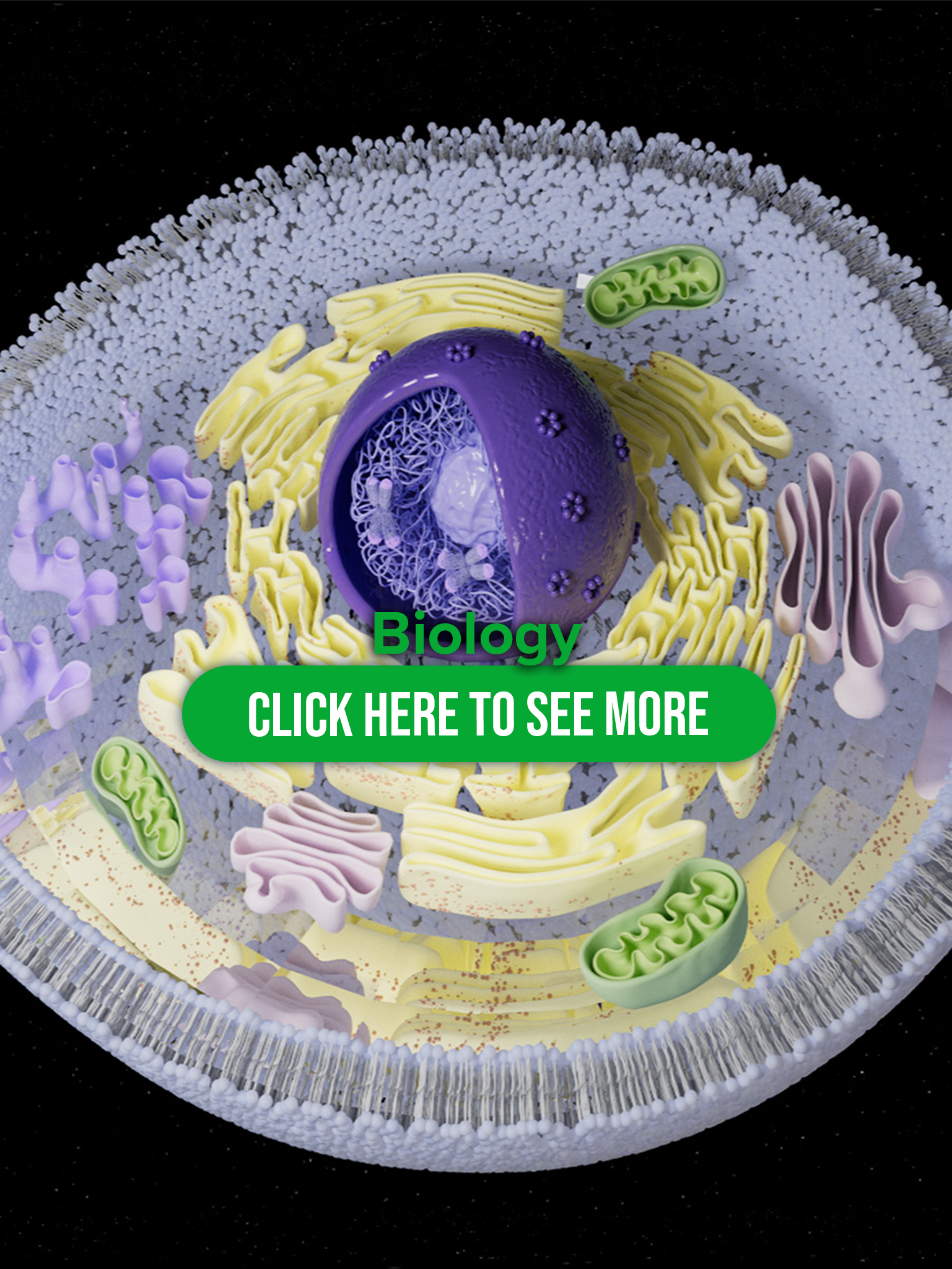
Description: AnatoMEE project is gamified digital toolkit to help undergraduate students better understand anatomy by transitioning from 2D to 3D learning.
01. UI element Design

Login Page Design

Error Page Design

Banner Design

Module Button Design
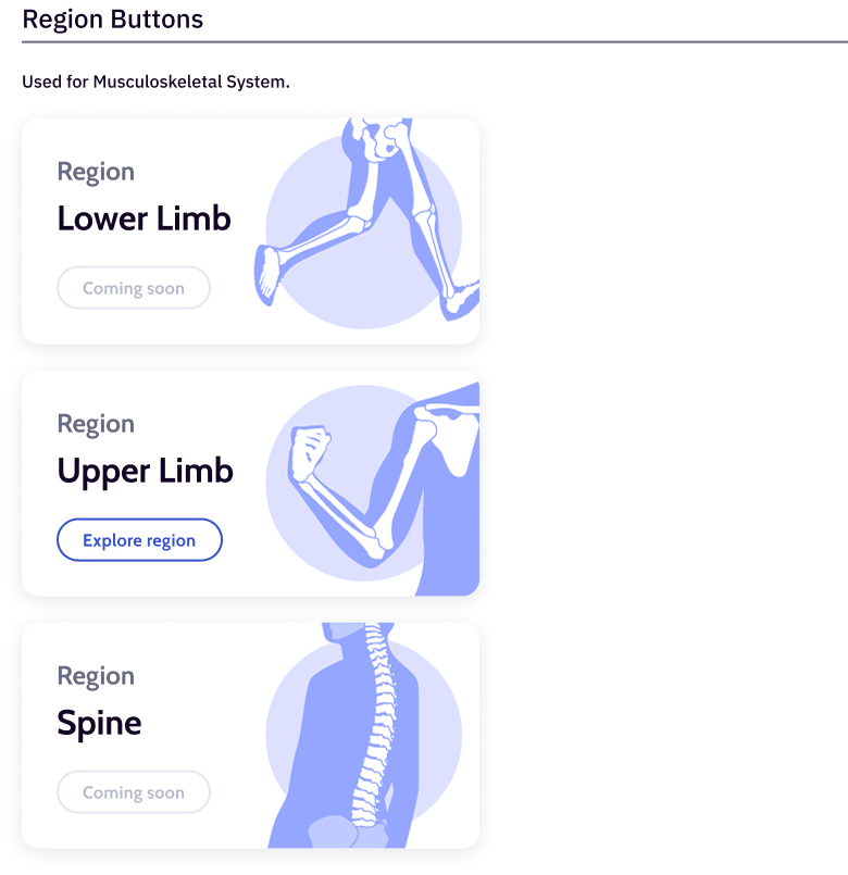
Module Button Design
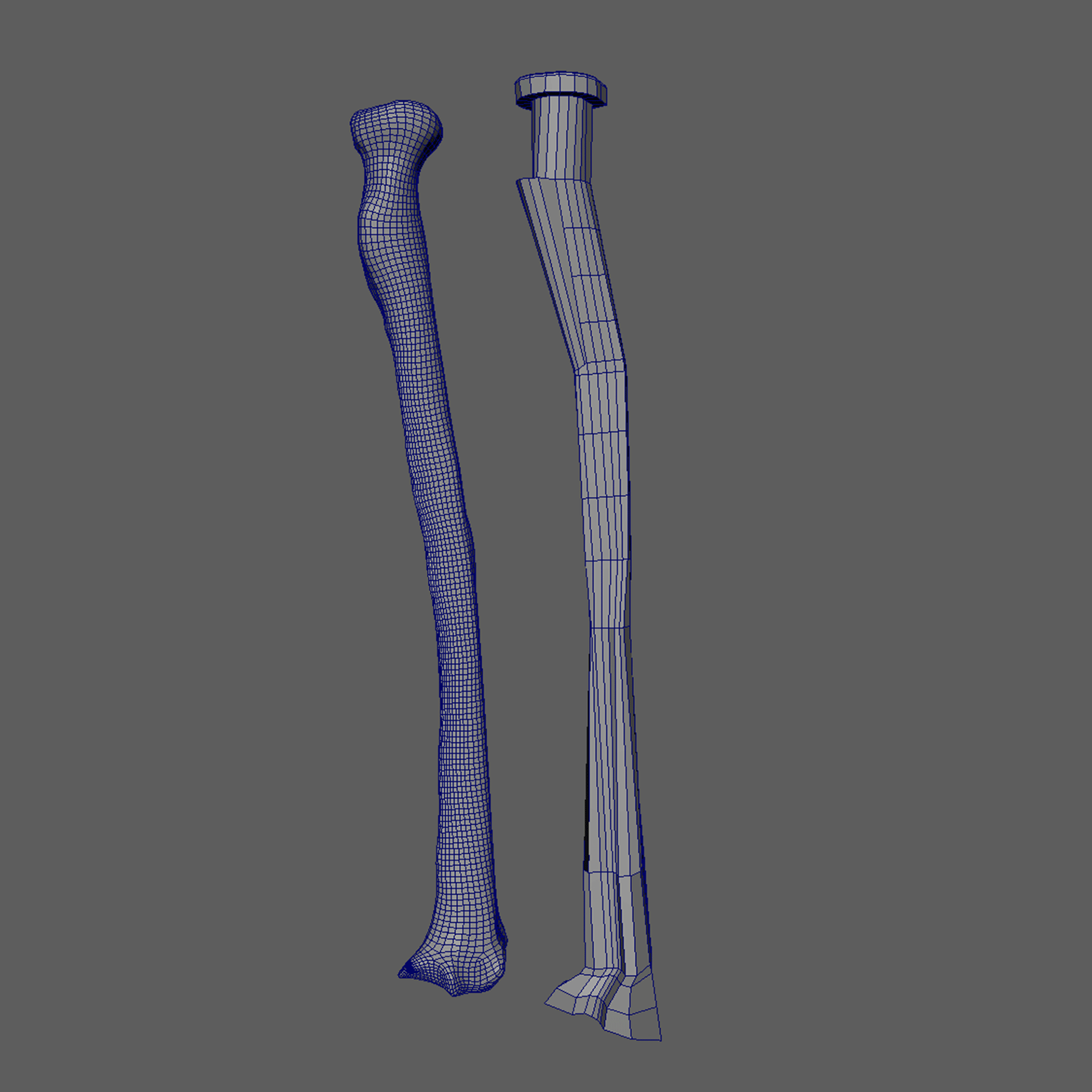
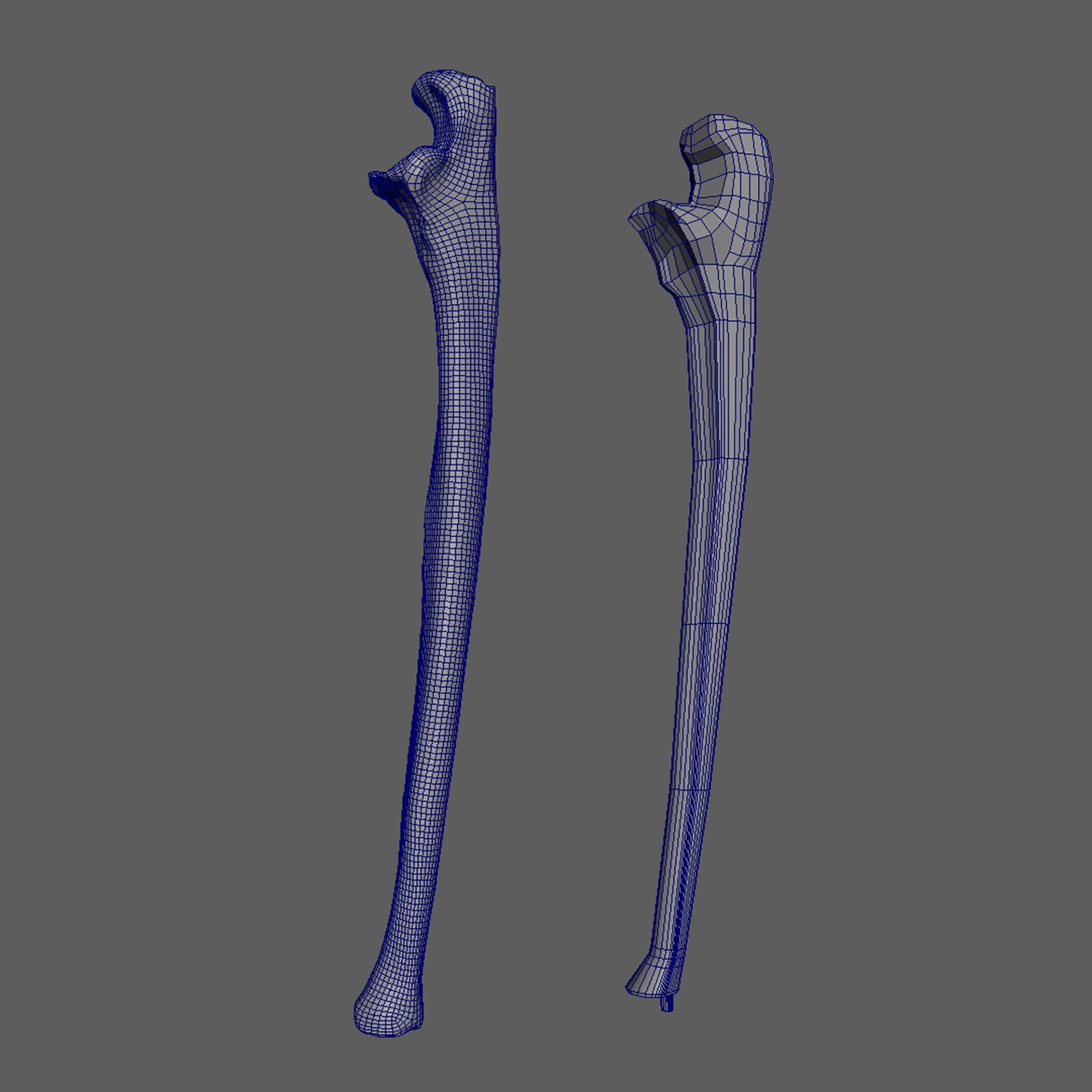
02. 3D Asset Creation (Realistic / Geometric ver.)
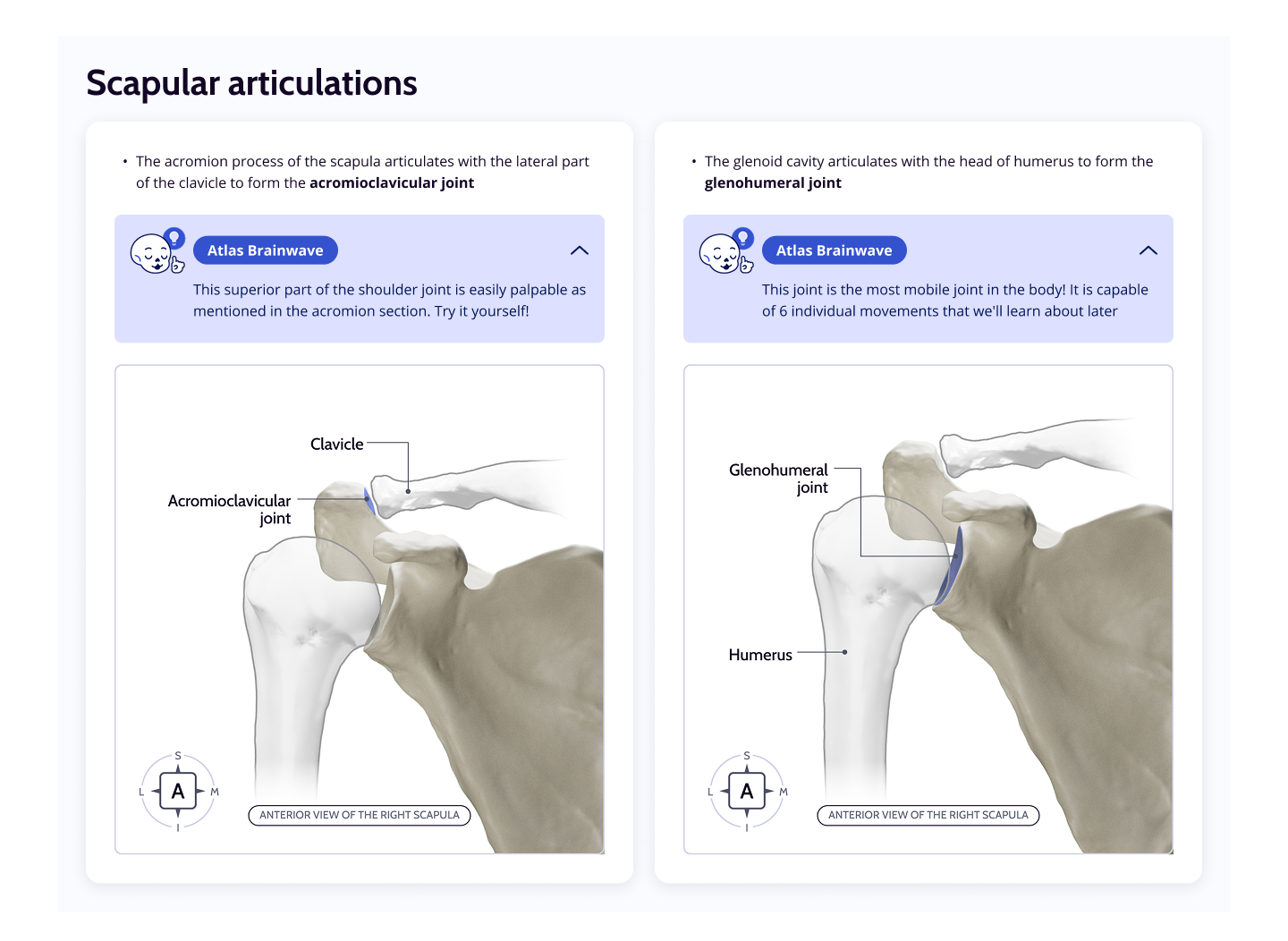
Scapular Articulations
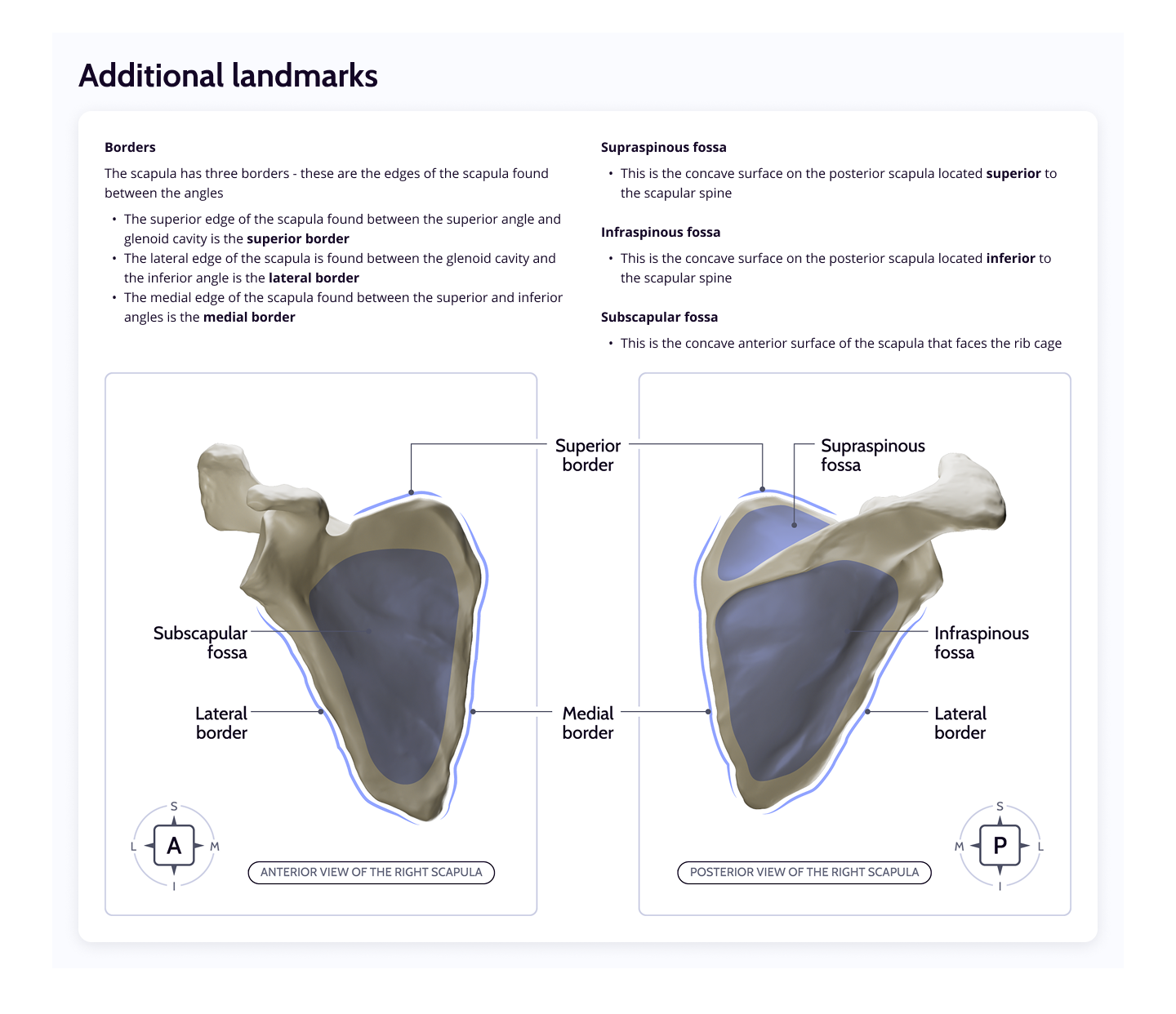
Scapula Landmarks
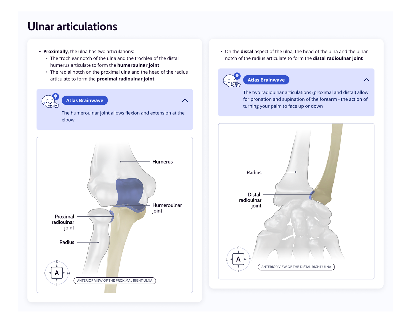
Ulnar Articulations
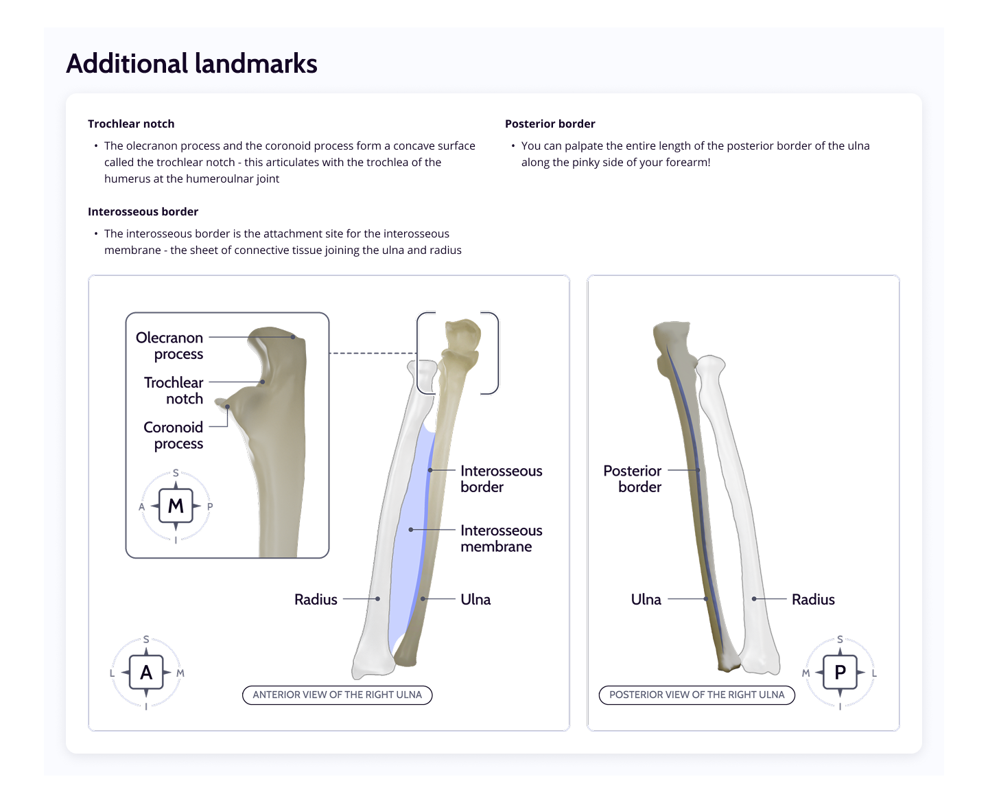
Ulna Landmarks
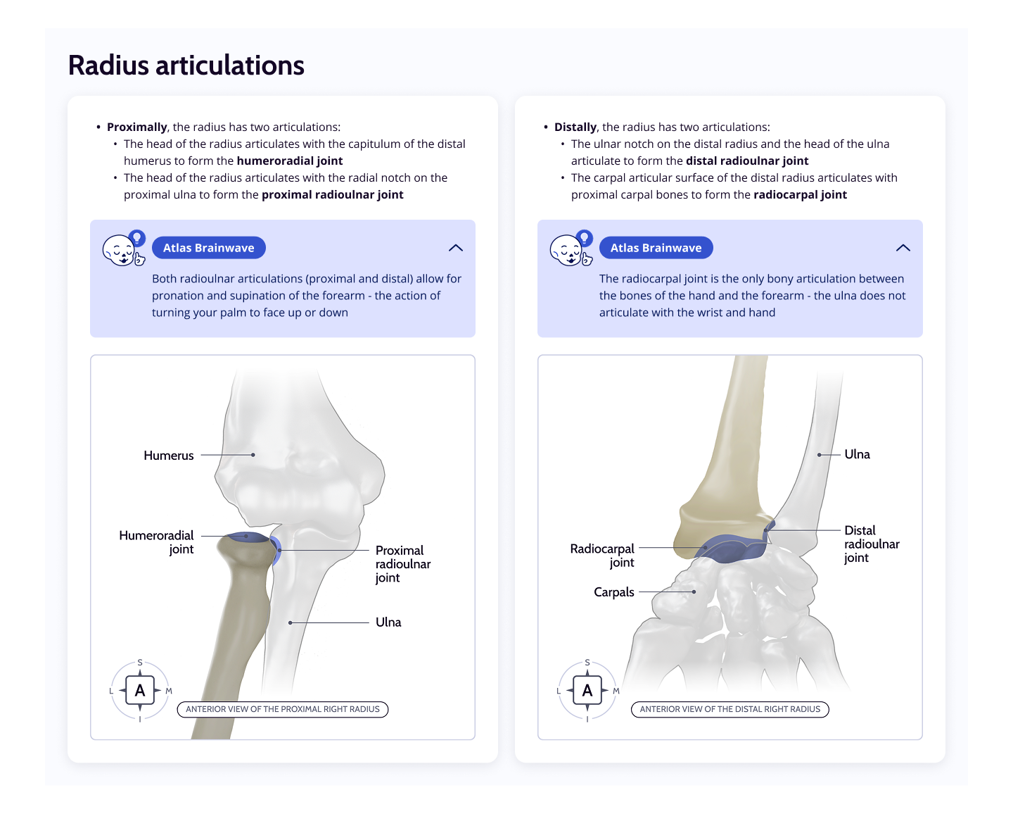
Radius Articulations
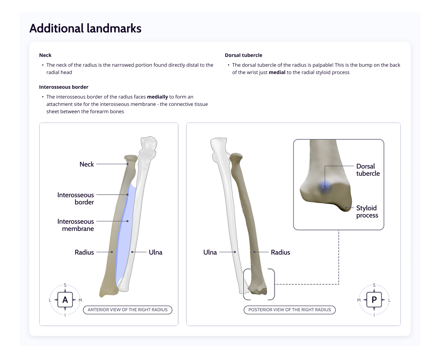
Radius Landmarks
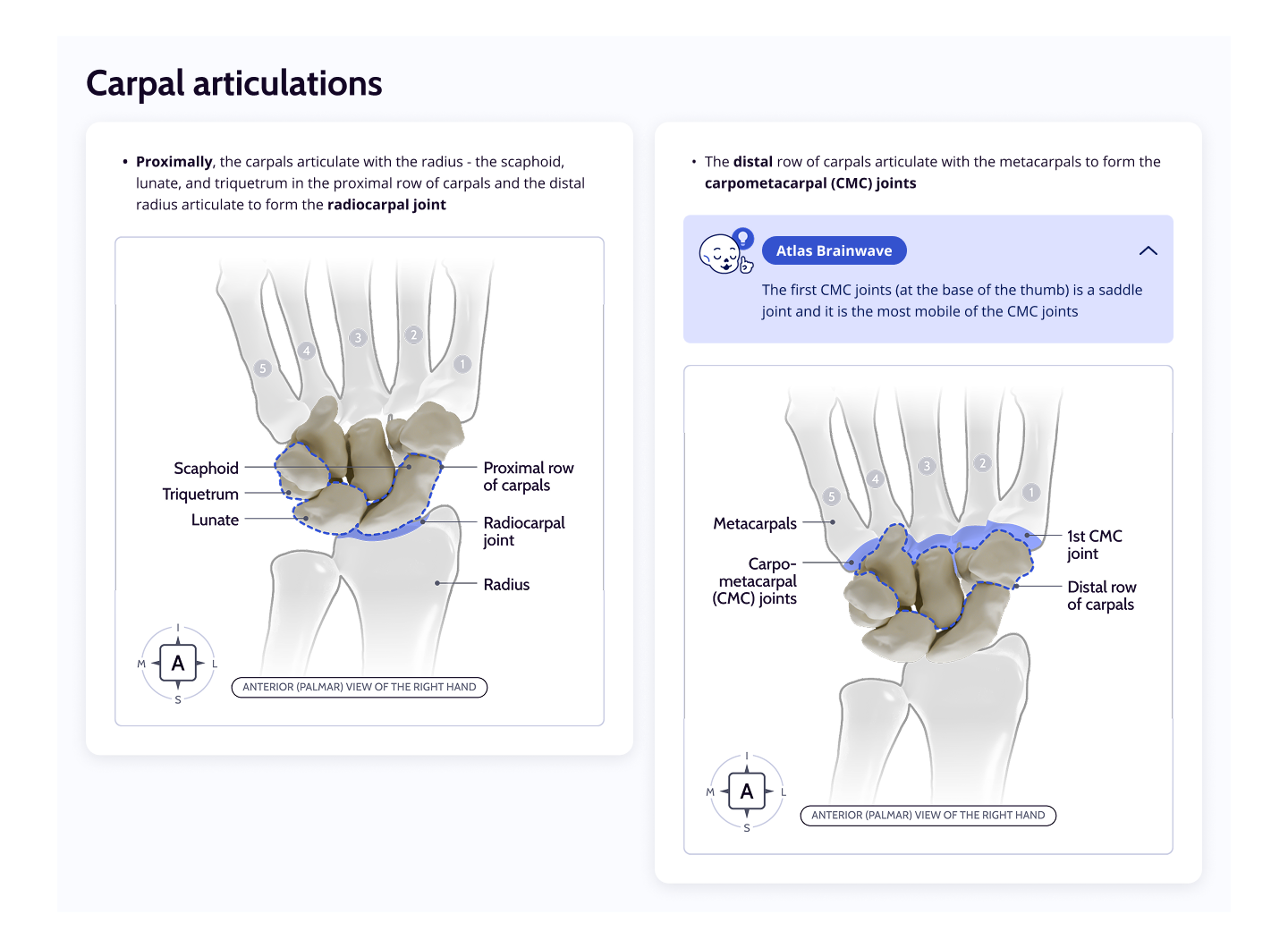
Carpal Articulations
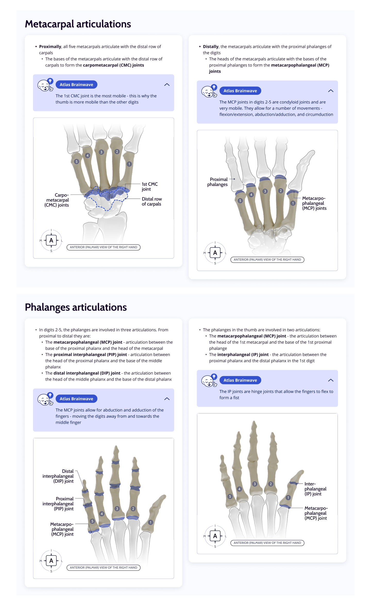
Metacarpal and Phalanges
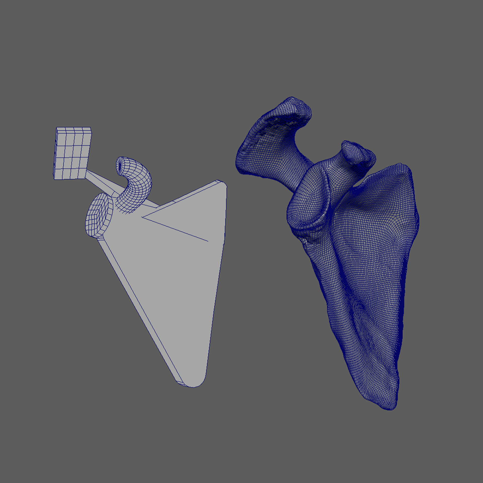
Scapula
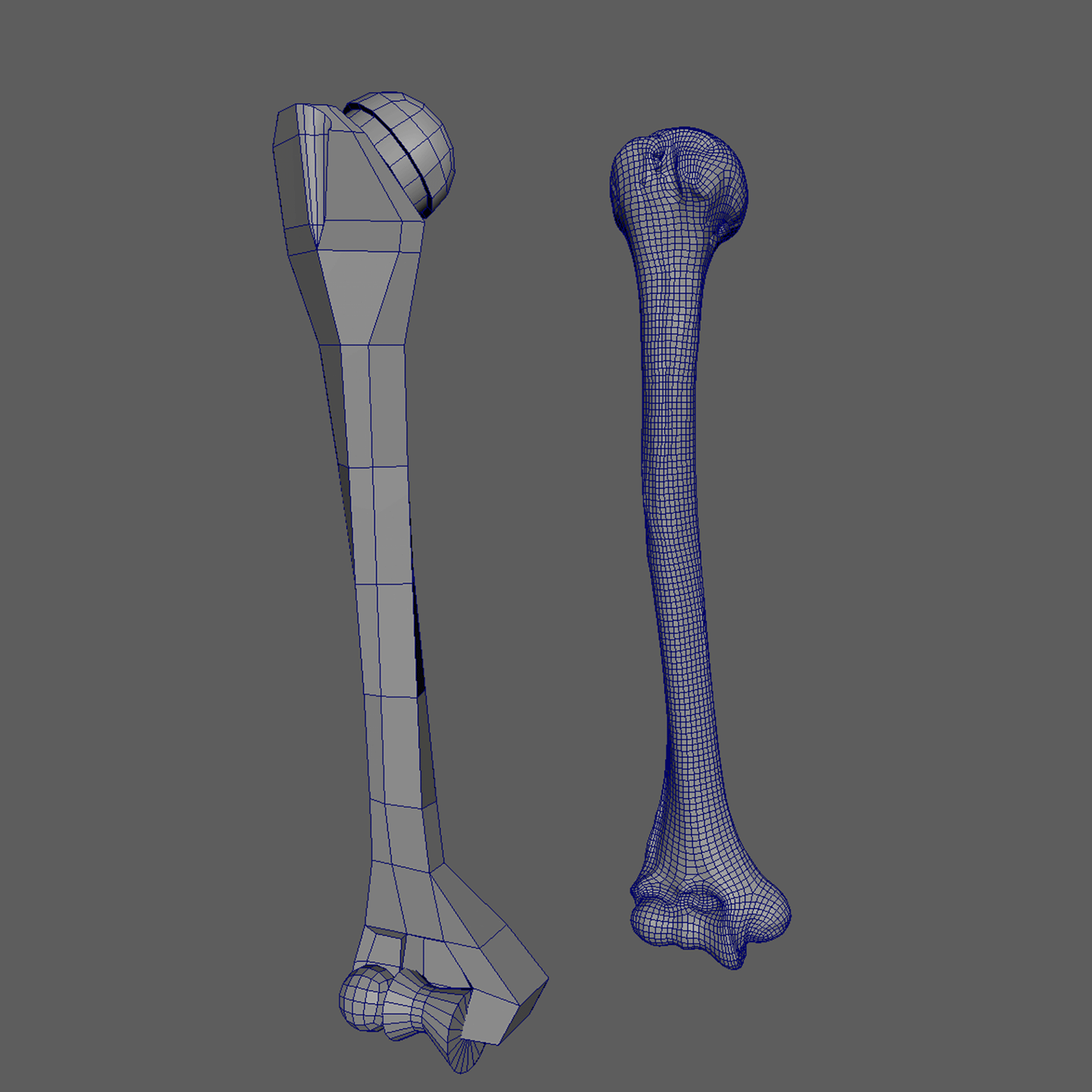
Humerus
Radius
Ulna
03. Animation Creation
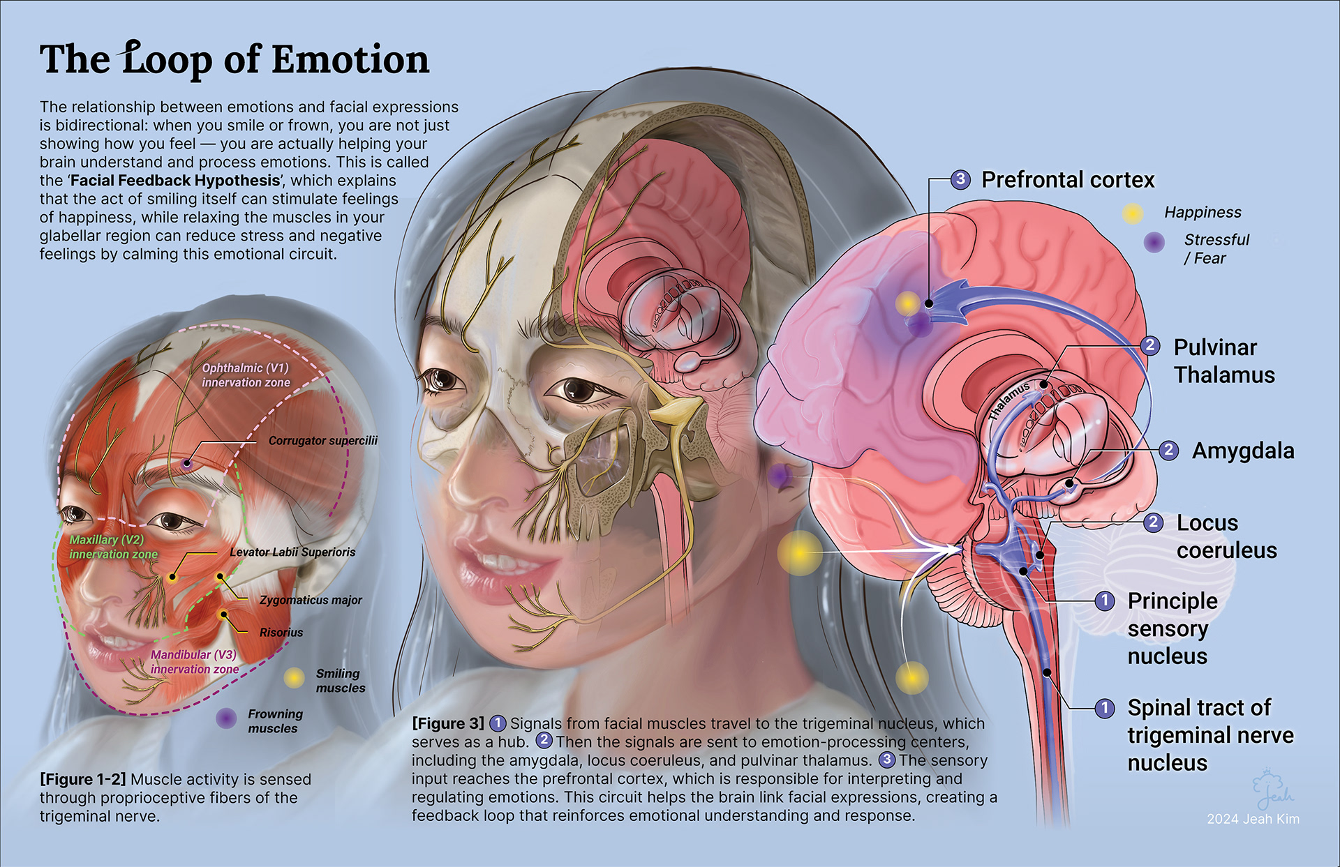
The Loop of Emotion (2024)
Media: Procreate, Adobe Illustrator
Description: This illustration explores the unseen dialogue between facial muscles and emotional experience. Inspired by research on how Botox affects not only facial movement but also emotion, the piece visualizes the Facial Feedback Hypothesis—the idea that our expressions are not just outward reflections of inner states but active participants in shaping them.
The brain doesn’t simply observe a smile or a furrowed brow; it senses muscle activity via proprioceptive fibers of the trigeminal nerve, interpreting these signals as emotional cues. From there, a cascade of neural communication unfolds: messages pass through the trigeminal nucleus to emotion-processing centers such as the amygdala, locus coeruleus, and pulvinar thalamus, eventually reaching the prefrontal cortex, where emotions are interpreted and regulated.
By revealing this hidden circuit, the piece makes visible how relaxing the glabellar region can ease stress or how the simple act of smiling can evoke joy—tiny movements with profound emotional consequences. This piece invites viewers not only to be informed but also to be uplifted—perhaps even to smile and feel a gentle sense of calm as they engage with it.
Selected as one of the 🏆 Top 12 finalists in the inaugural Art Neureau 2025 exhibition,
'Sensing the Unseen' [Link to the Virtual Exhibition] 🎉🥳🎨🖌️
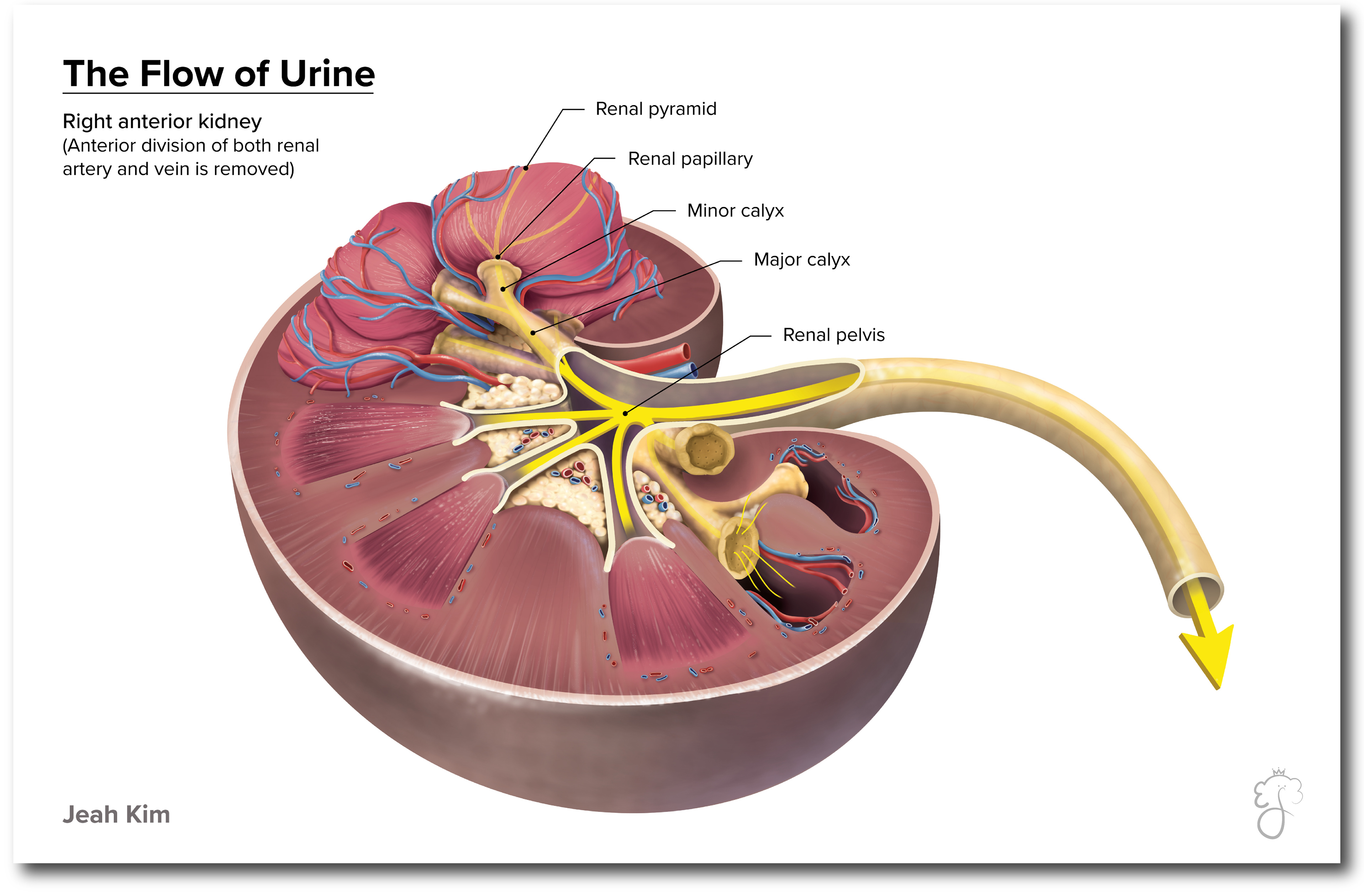
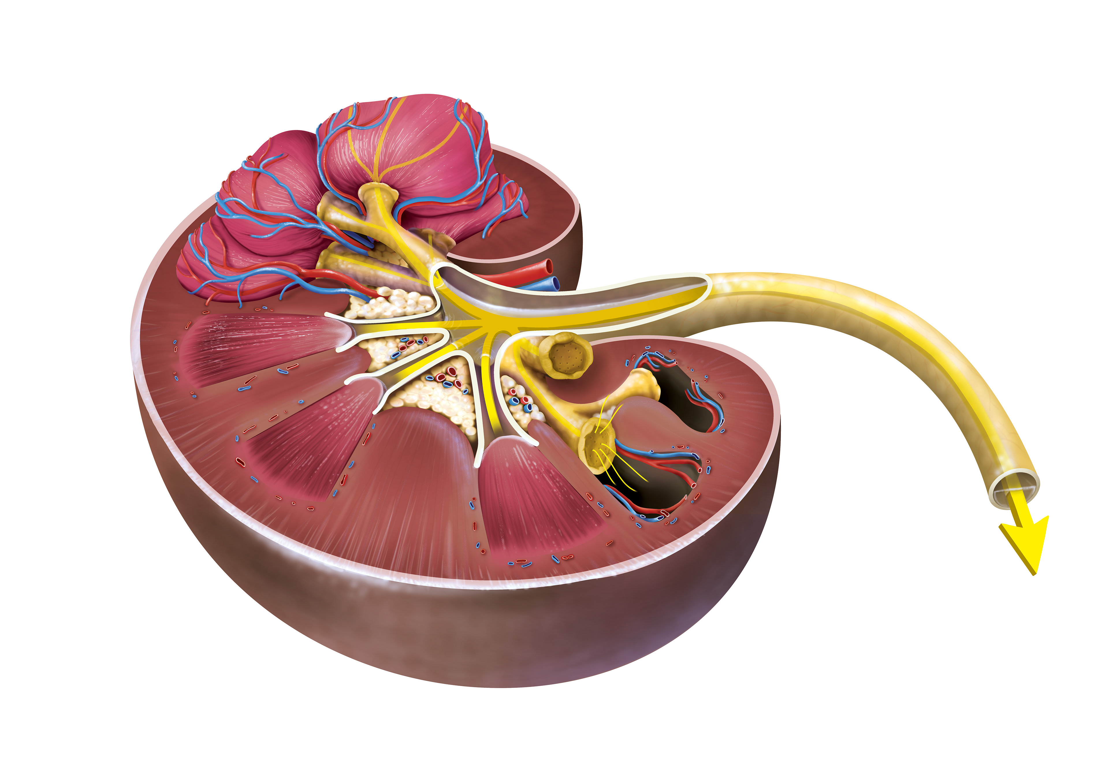
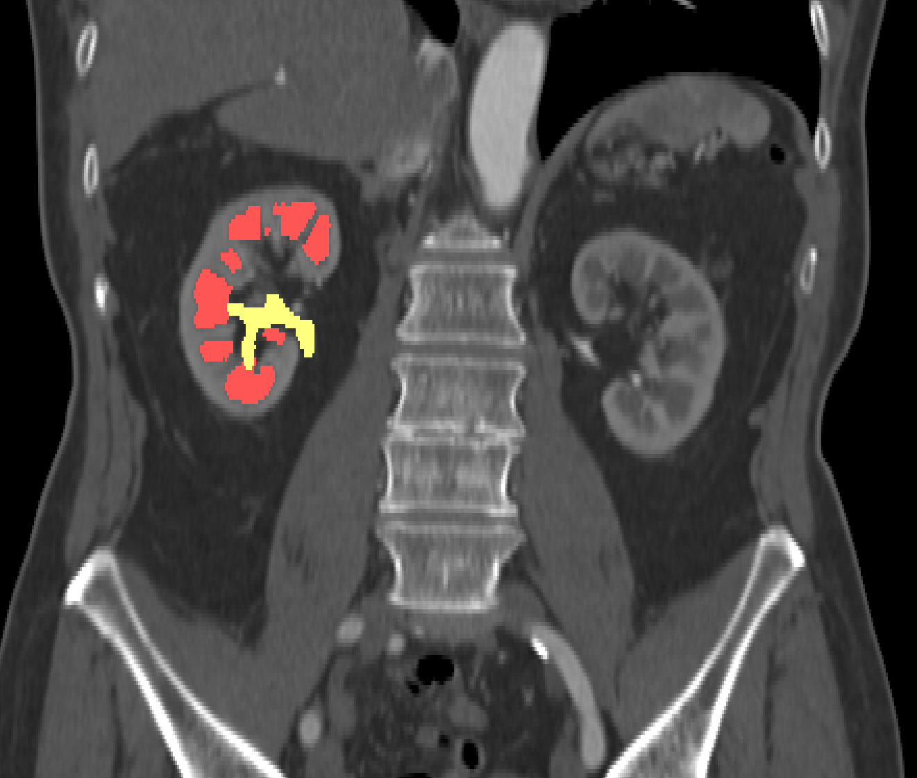
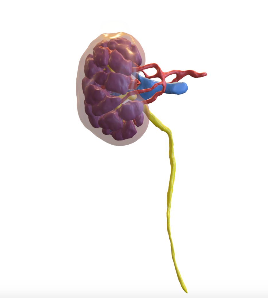
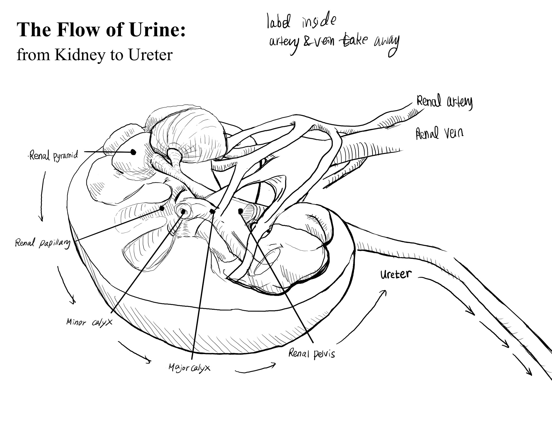
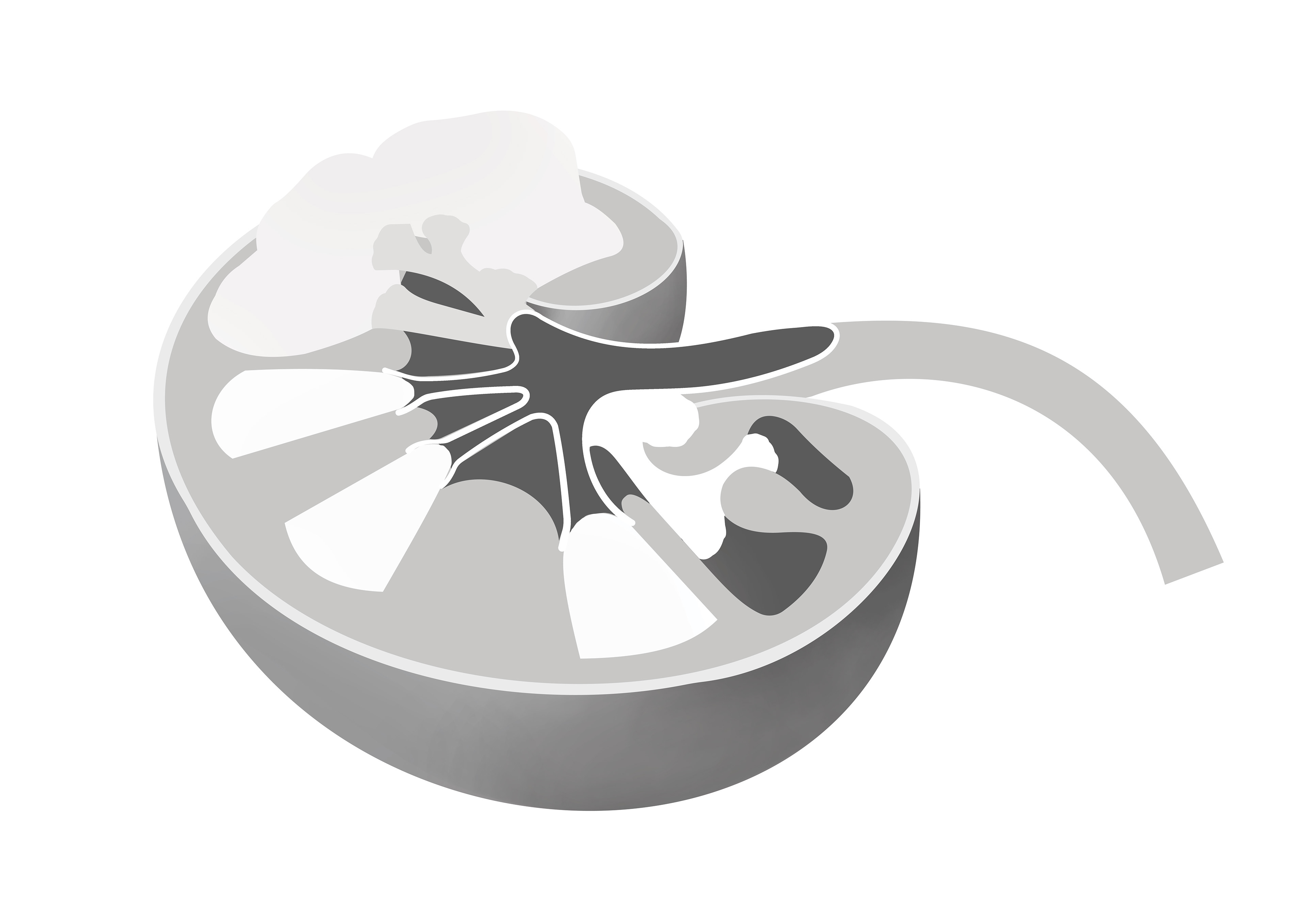
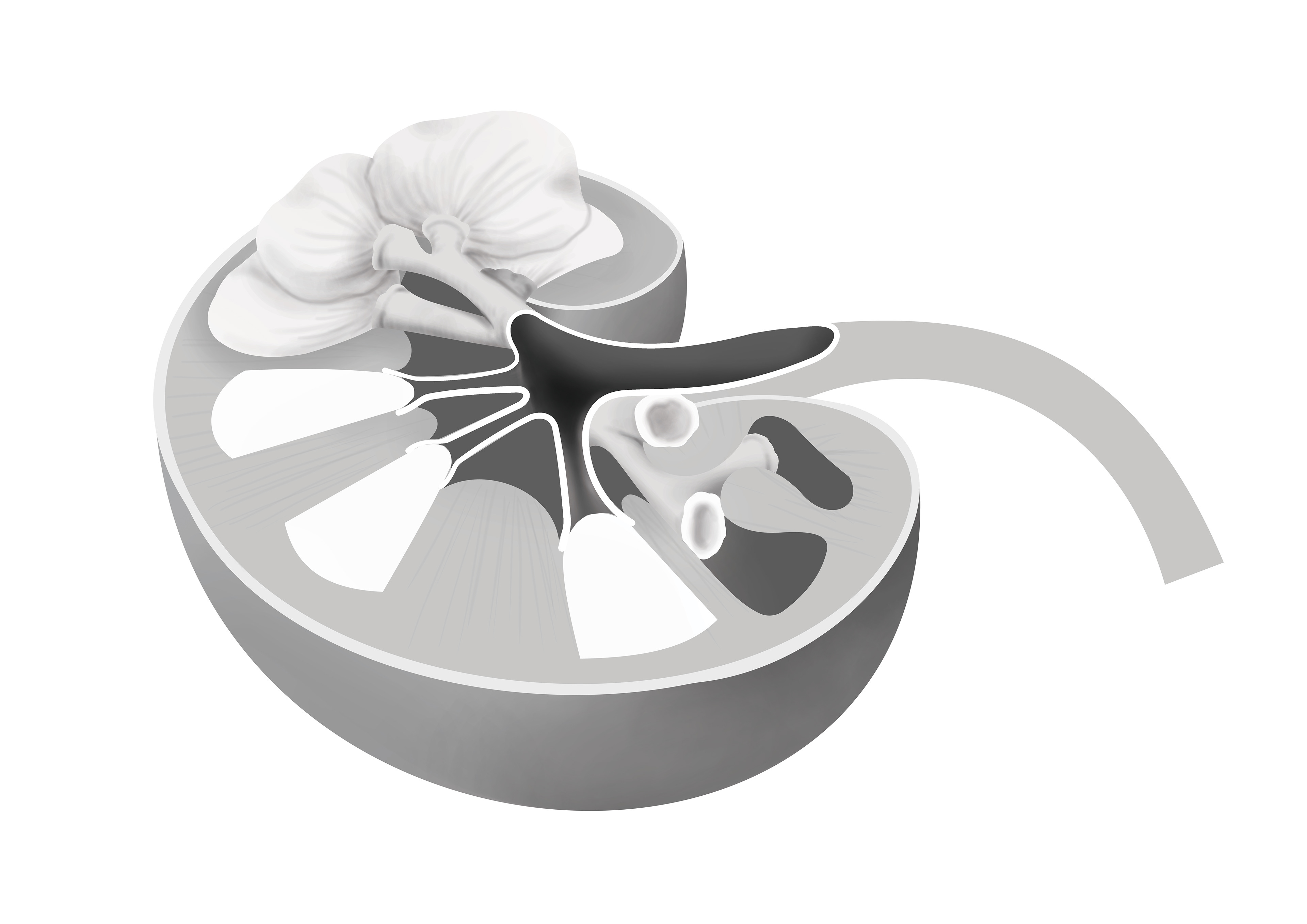
The Flow of Urine (2024)
Media: Adobe Photoshop
Description: Spotted a unique view, but also effectively showing the sequential process of urine traveling from the renal medulla into the ureter. Labels are harmonized to the drawing and kept in order which makes reader easier to follow the flow of urine. As it displays 3D, 2D (plane cut) and removed versions of the medulla, readers can understand the three-dimensional structure of the medulla filling the kidney and observe how renal vessels surround each pyramid. Specifically, by showing the part with the pyramid removed, it depicts the terminal ends of the calyces, allowing readers to visualize renal tubules collected in the renal papillary region.
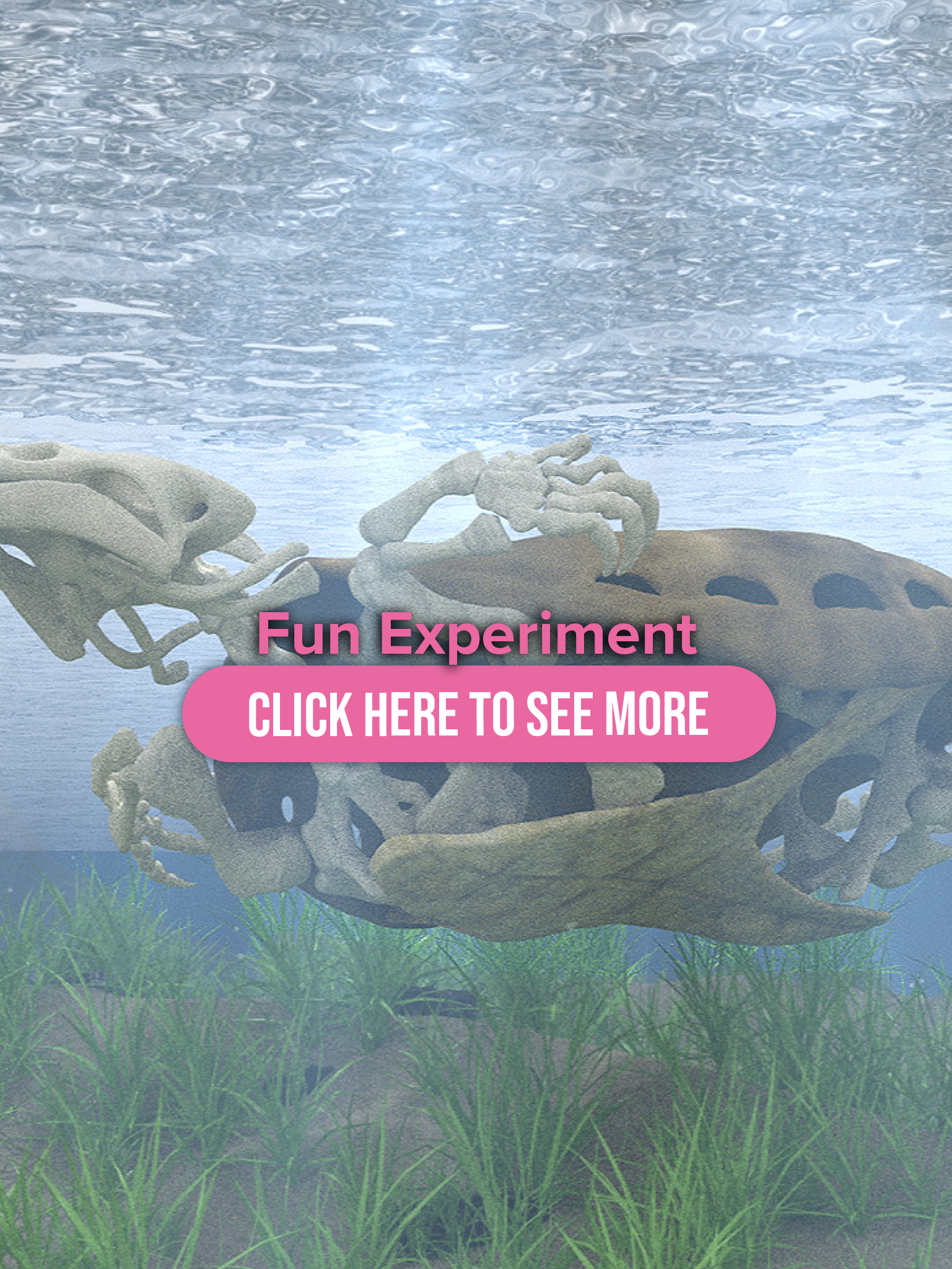
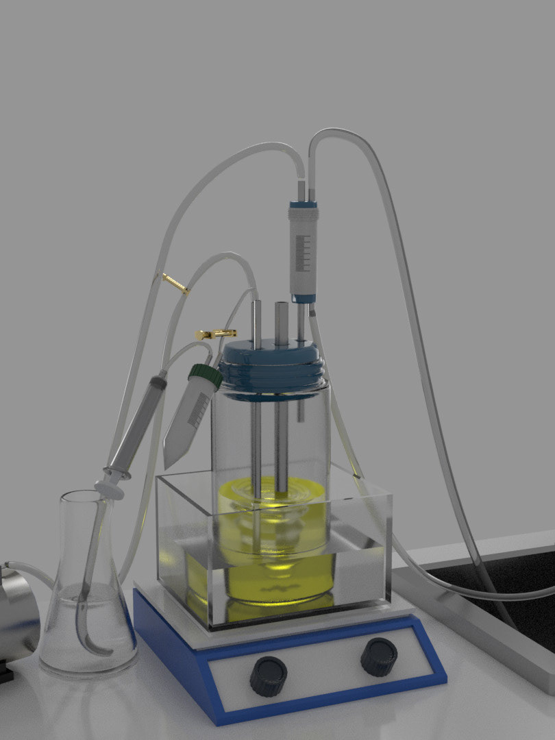
Data Sculpt 1
Data Sculpt 2
Sketch 1
Sketch 2
Sketch 3
Carbon Dust Technique (2023)
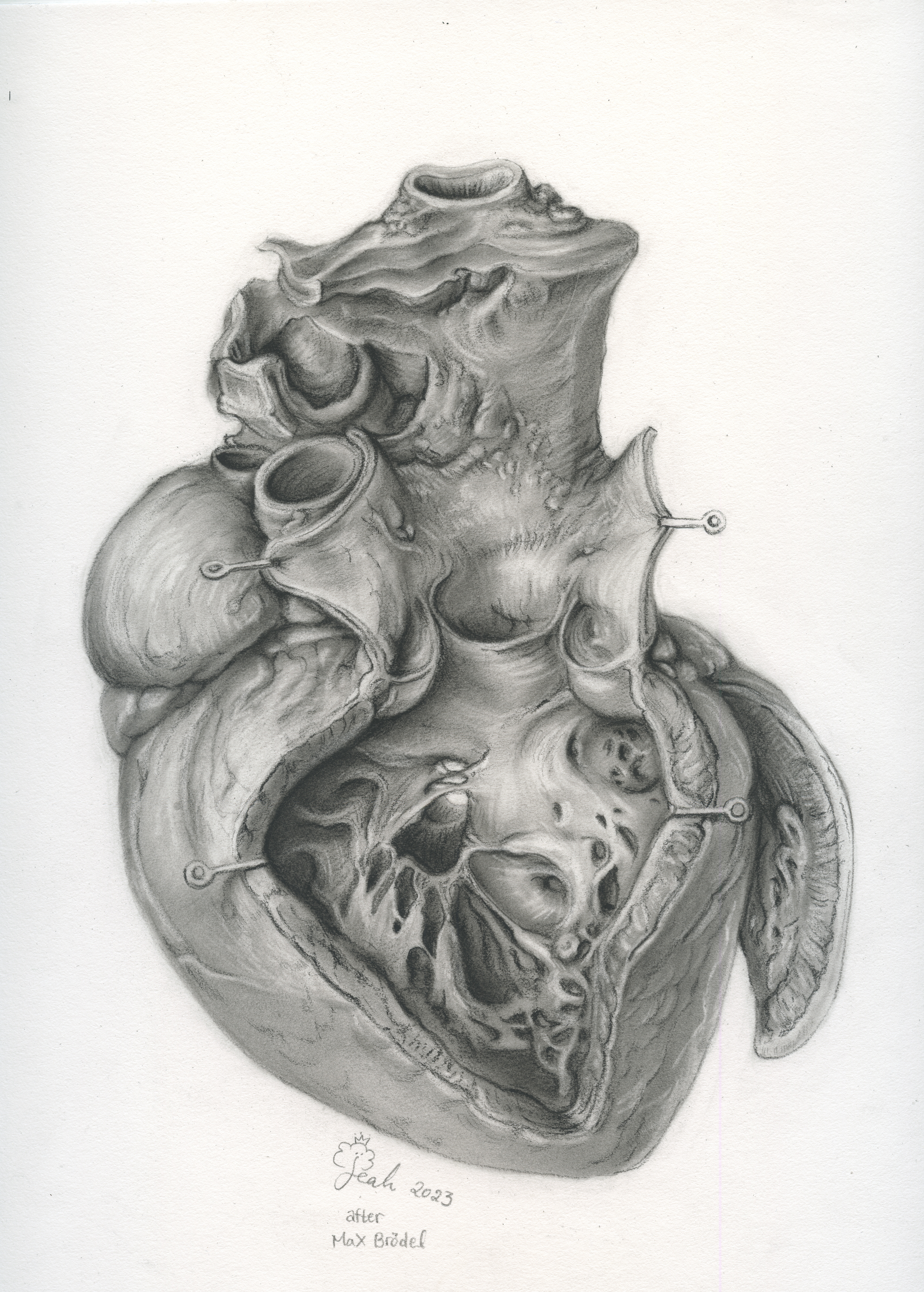
Heart after Max Brödel
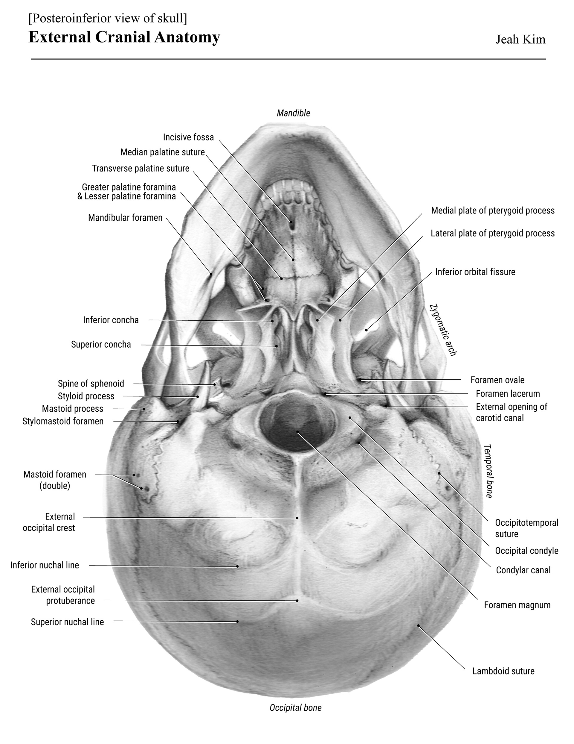
External Cranial Anatomy
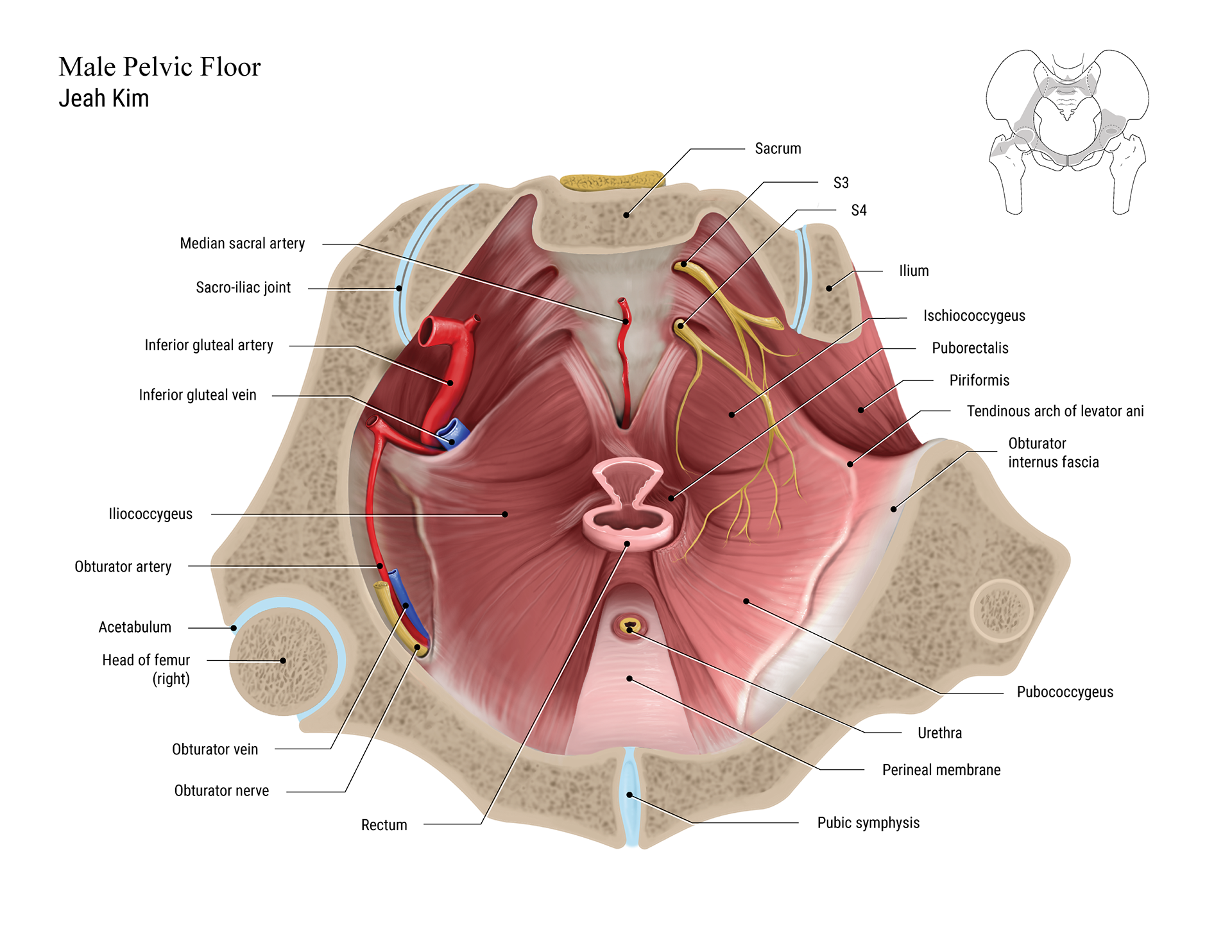
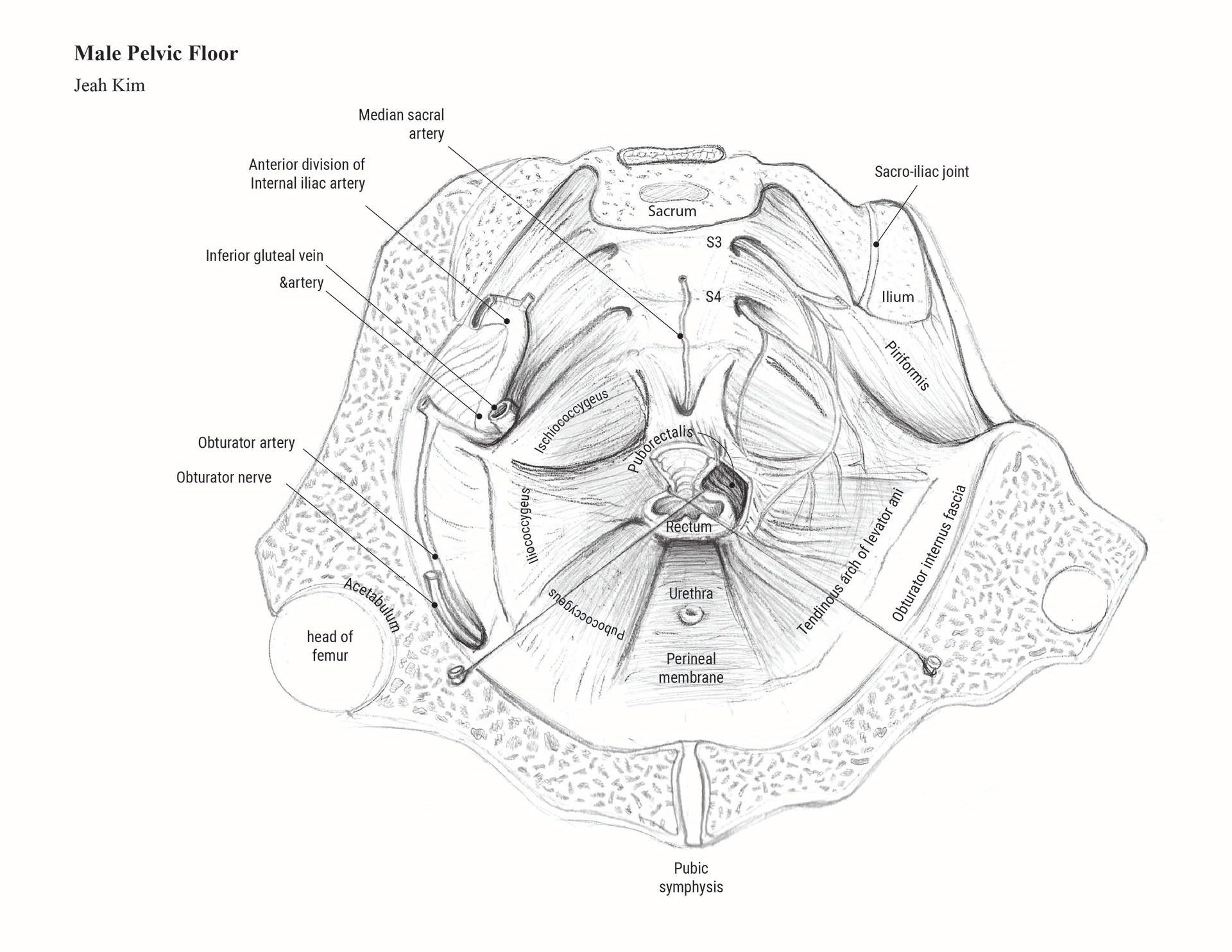
Male Pelvic Floor (2023)
Media: Adobe Photoshop
Description: Specimen drawing at Grant's Museum, Toronto
Targeted for the students who study the pelvic anatomical structure from the actual specimen. To help readers understand the unique cut of the specimen, I added a simple graphic about its orientation at the upper right corner.
Male Pelvic Floor_Colour
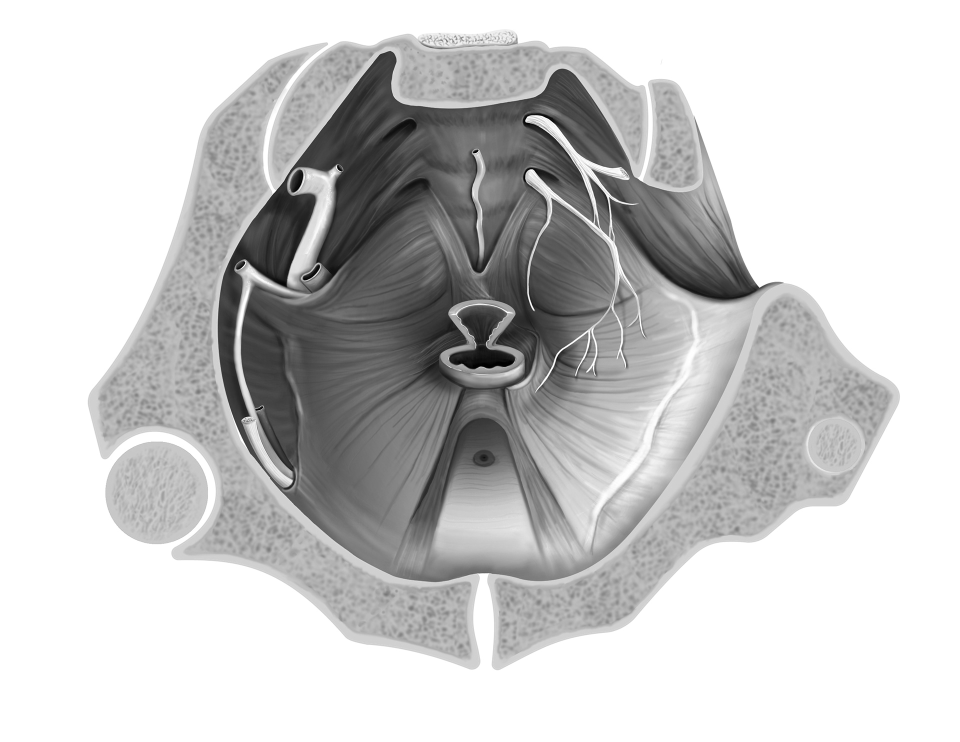
Male Pelvic Floor_Grey Scale
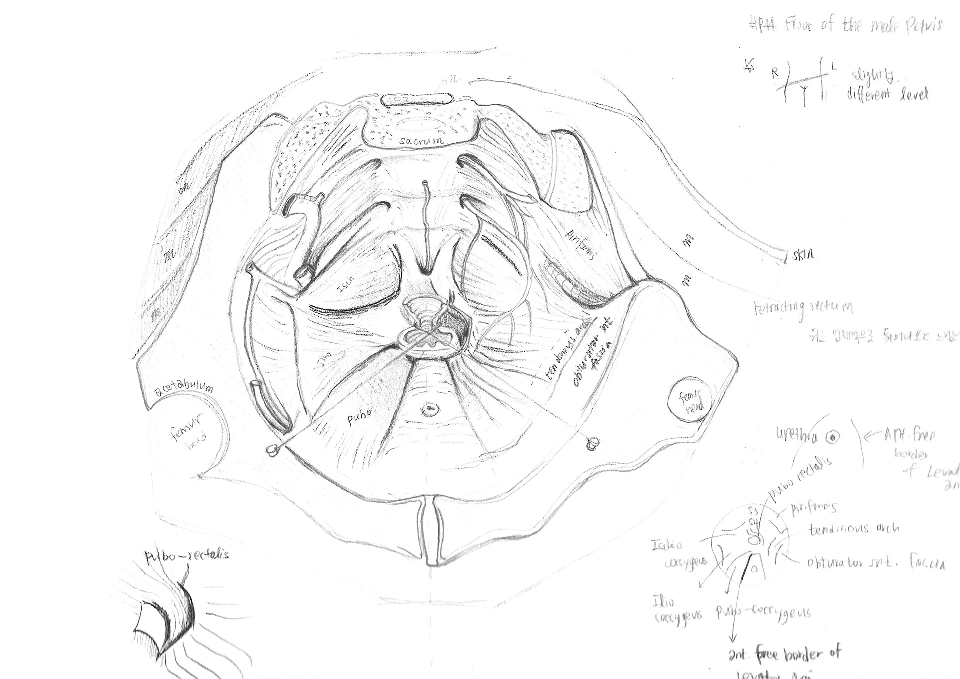
Male Pelvic Floor_Sketch 1

Male Pelvic Floor_Sketch 2
Male Pelvic Floor_Sketch 3
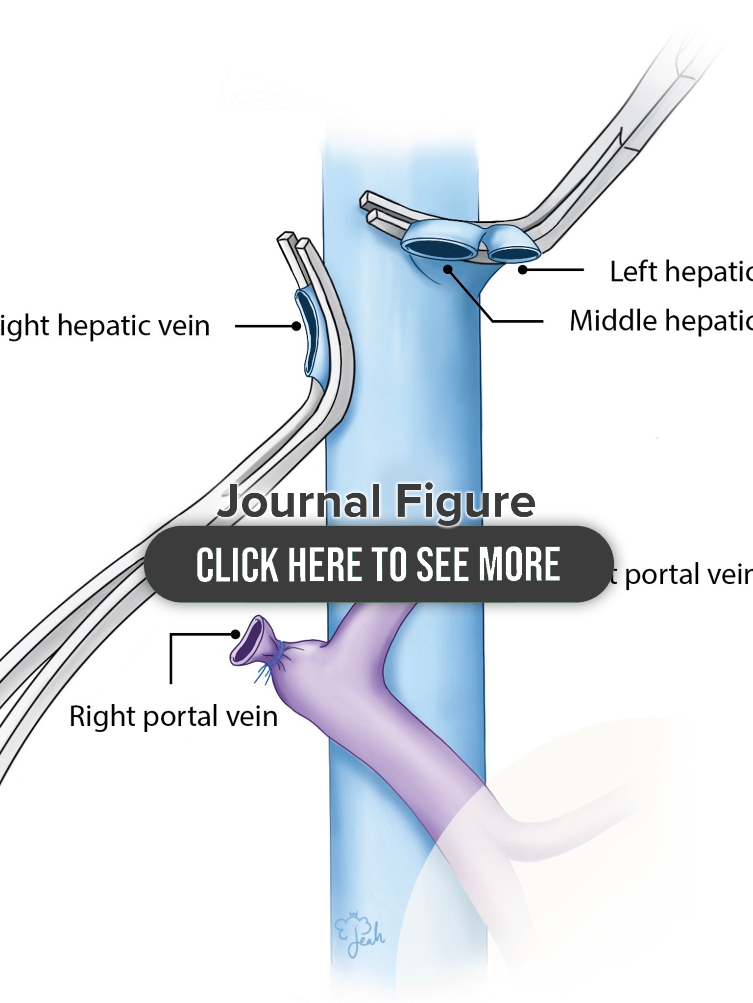
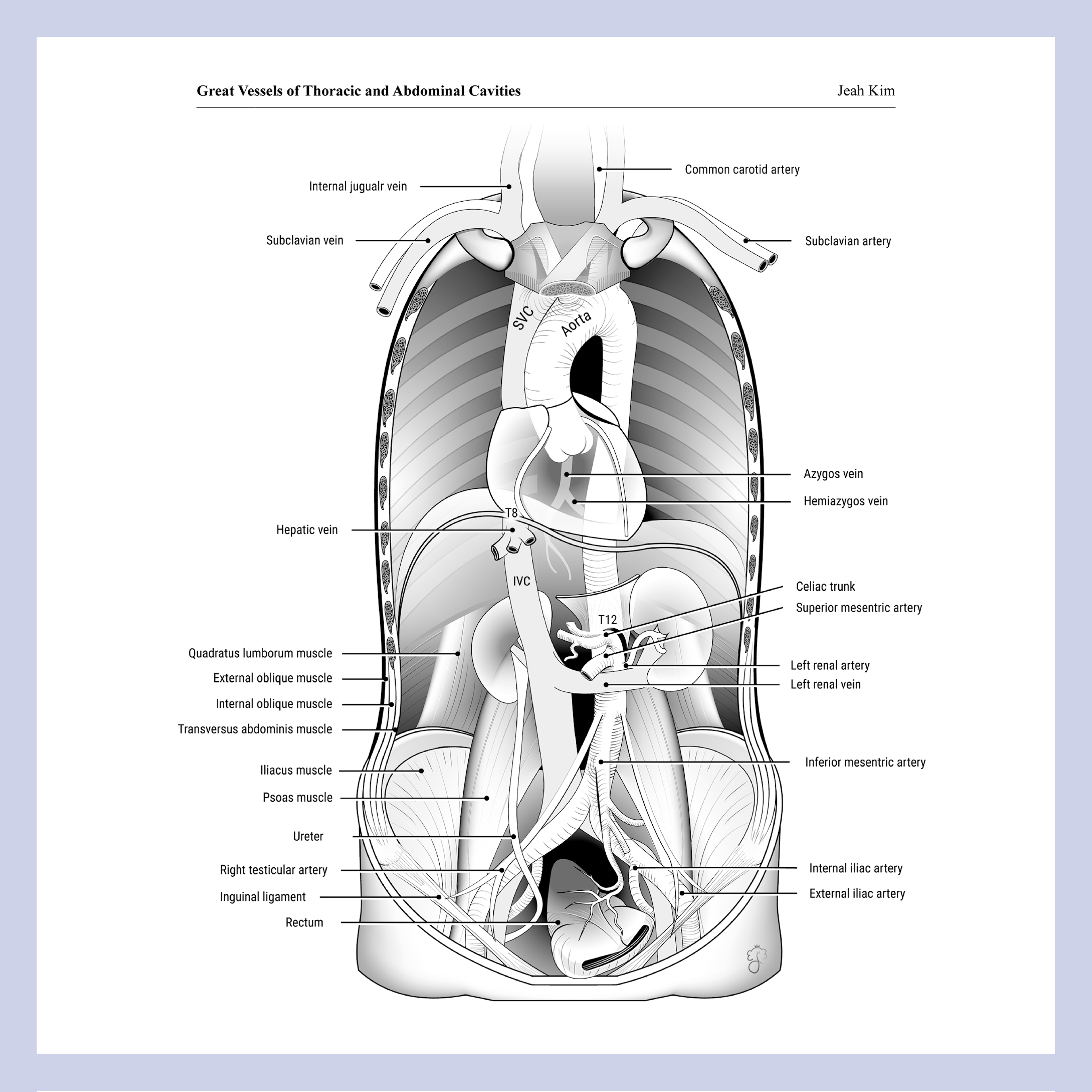
Great Vessels of Thoracic and Abdominal Cavities (2023)
Media: Adobe Illustrator
Description: Specimen drawing at Grant's Museum, Toronto
Specimen Drawing at Seoul St.Mary's Hospital, Korea (2022)
Media: Pencil, Procreate (coloured)
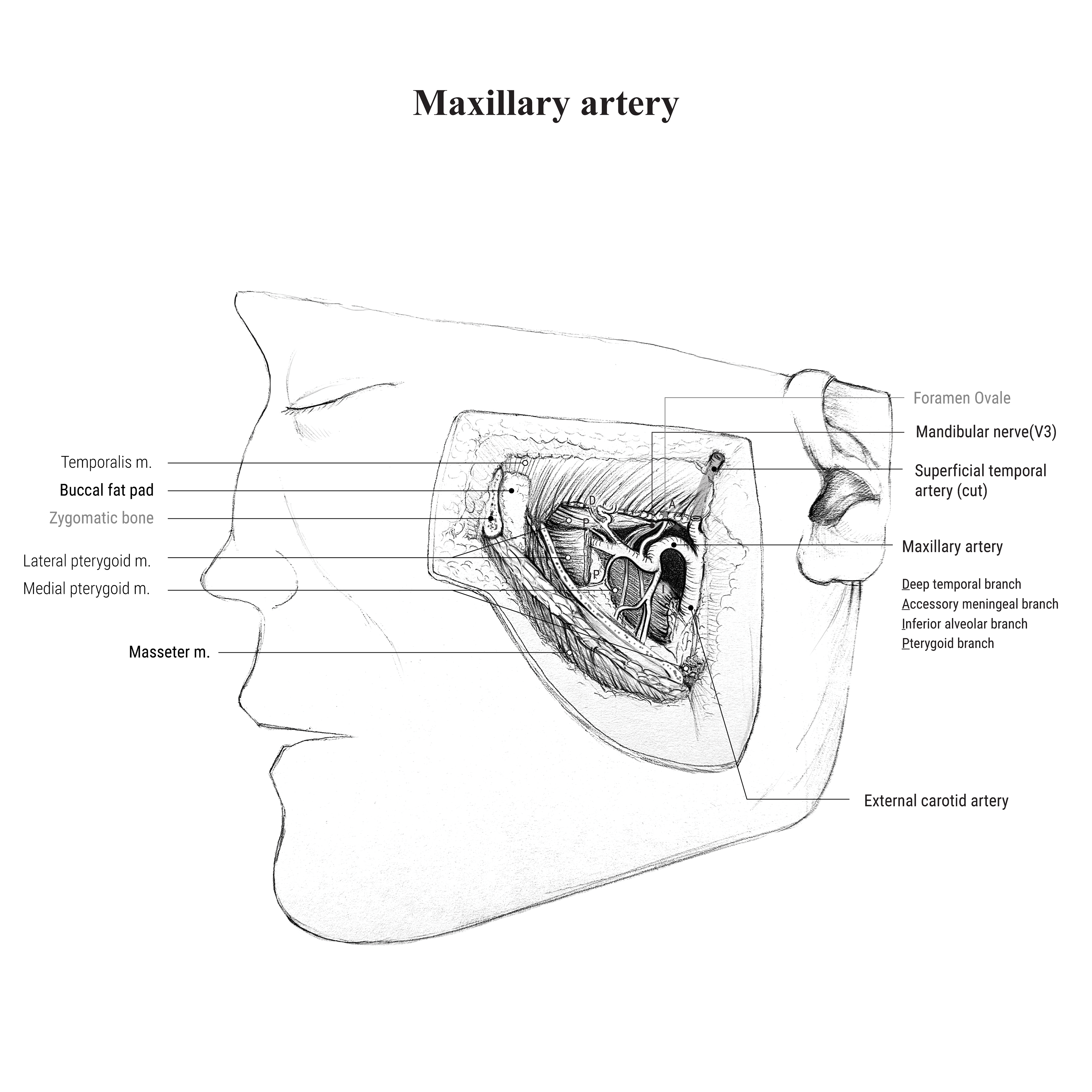
Maxillary Artery

Knee Joint
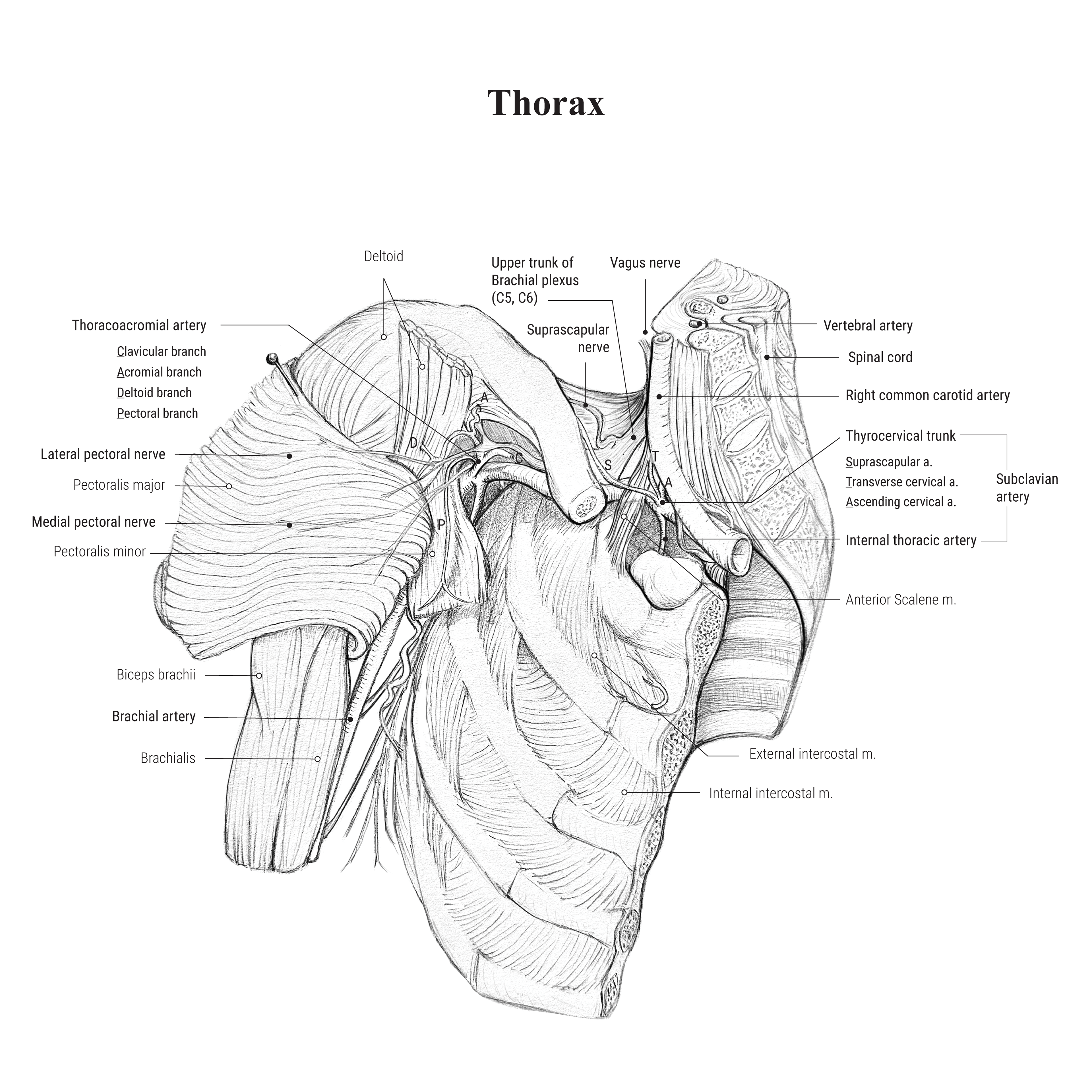
Thorax
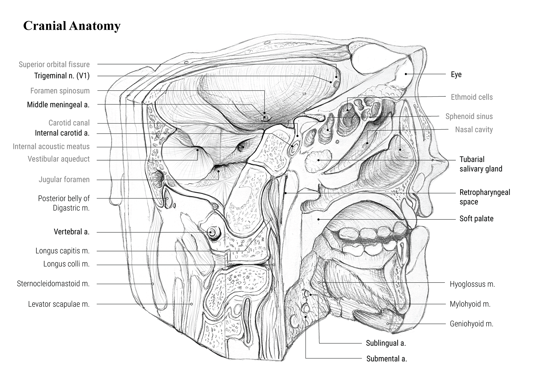
Cranial Anatomy
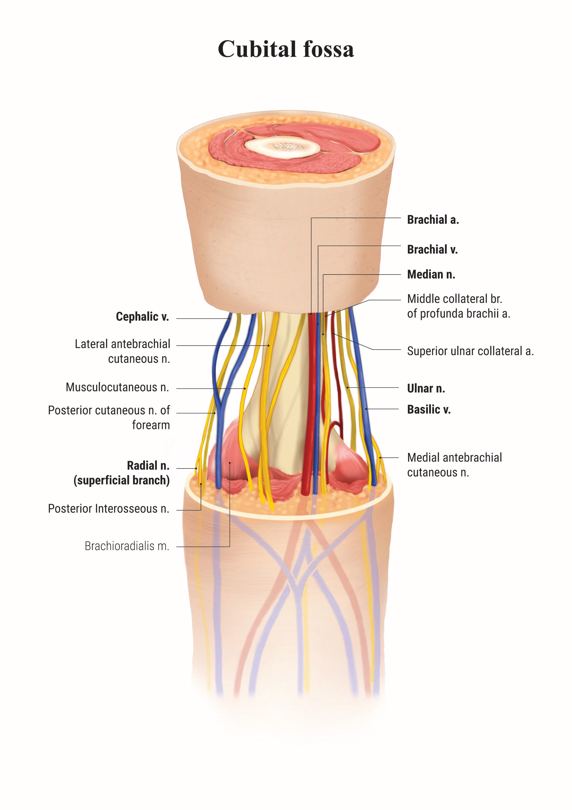
Cubital Fossa_Anterior view
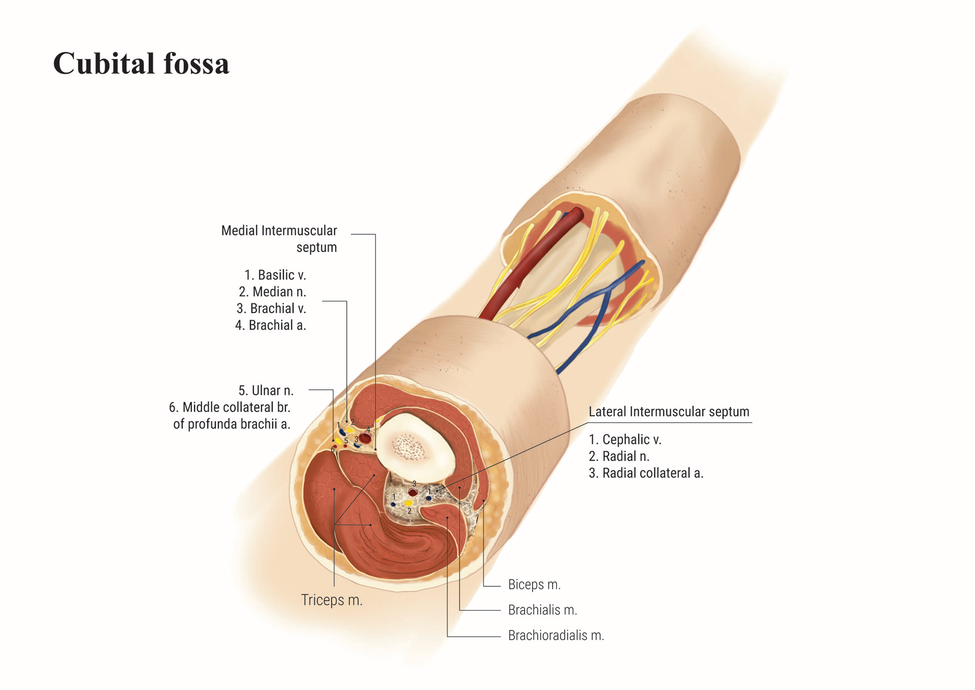
Cubital Fossa_Right Arm
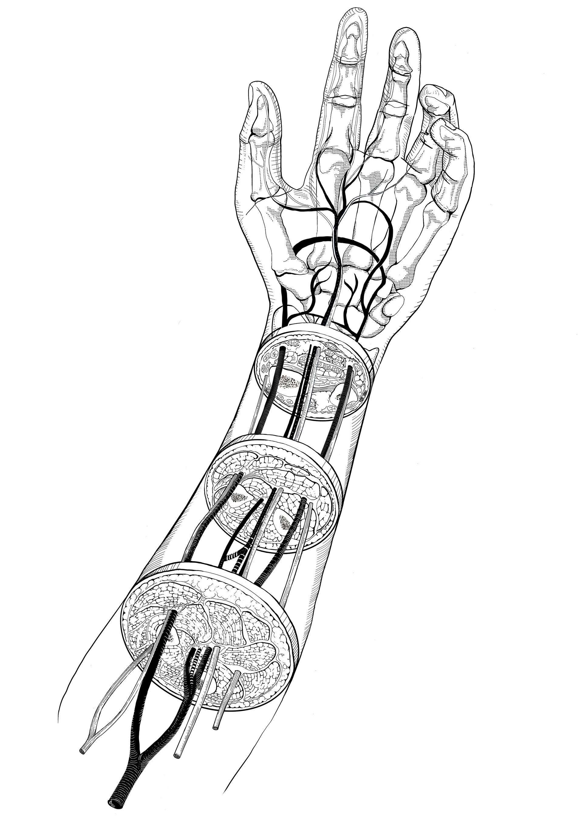
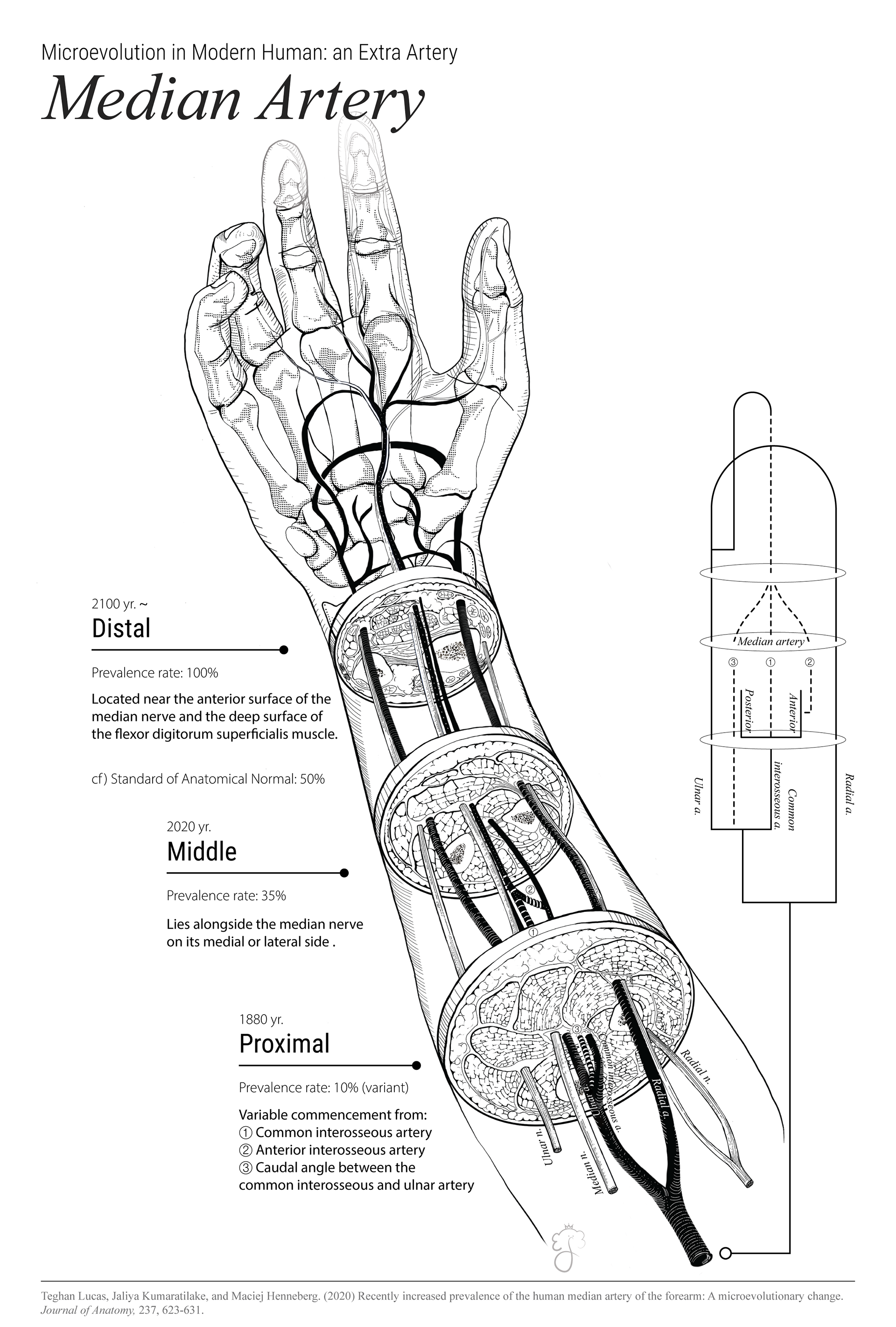
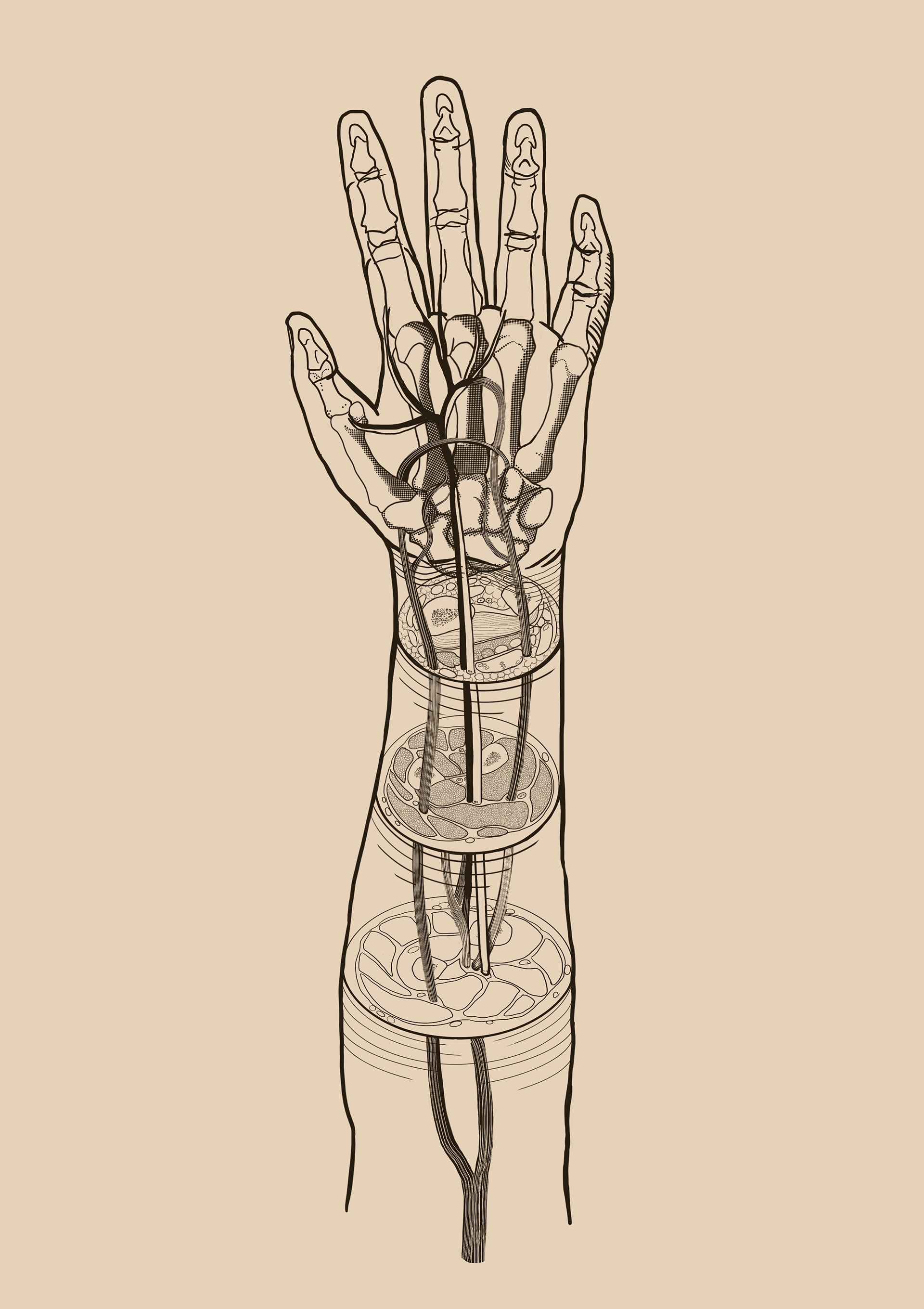
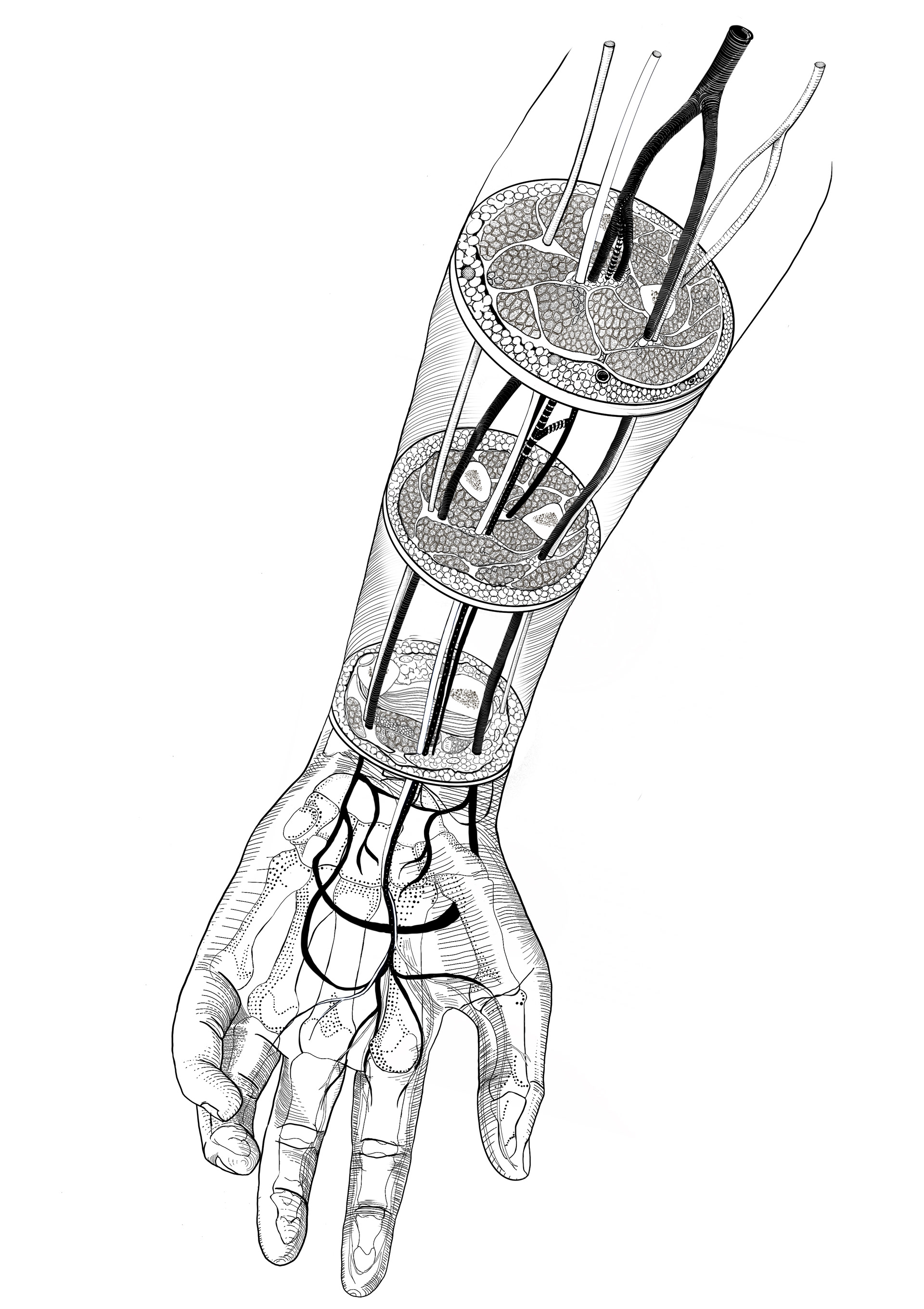
Microevolution in Modern Human: an Extra Artery, 'Median Artery' (2022)
Media: Procreate, Adobe Illustrator
Description: The median artery was traditionally a structure that regressed around the 8th week of pregnancy when the brachial and ulnar arteries formed. However, it is now found in about one-third of modern individuals, emerging as anatomical evidence that humans are continuously evolving.
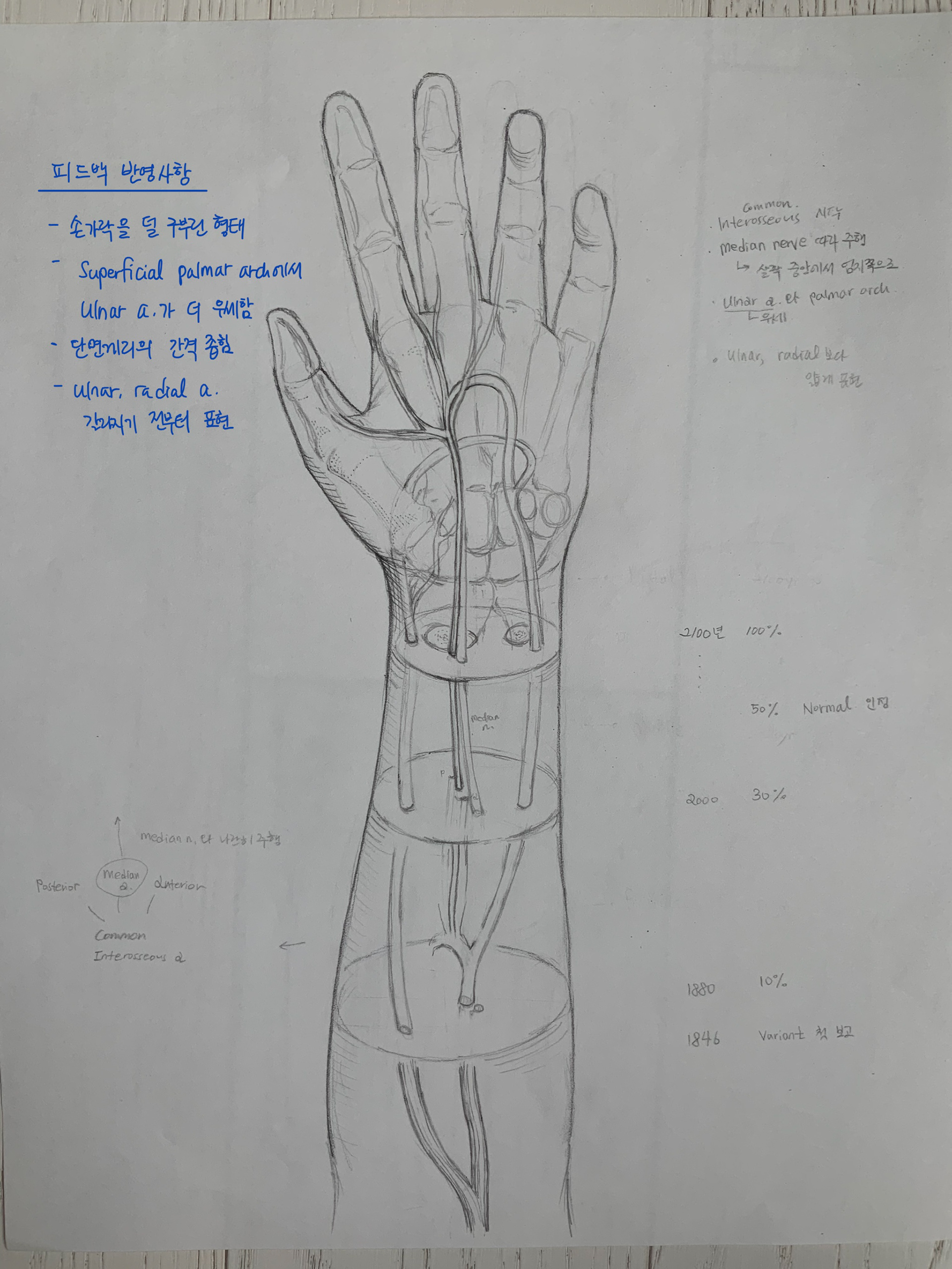
Median Artery_Sketch1
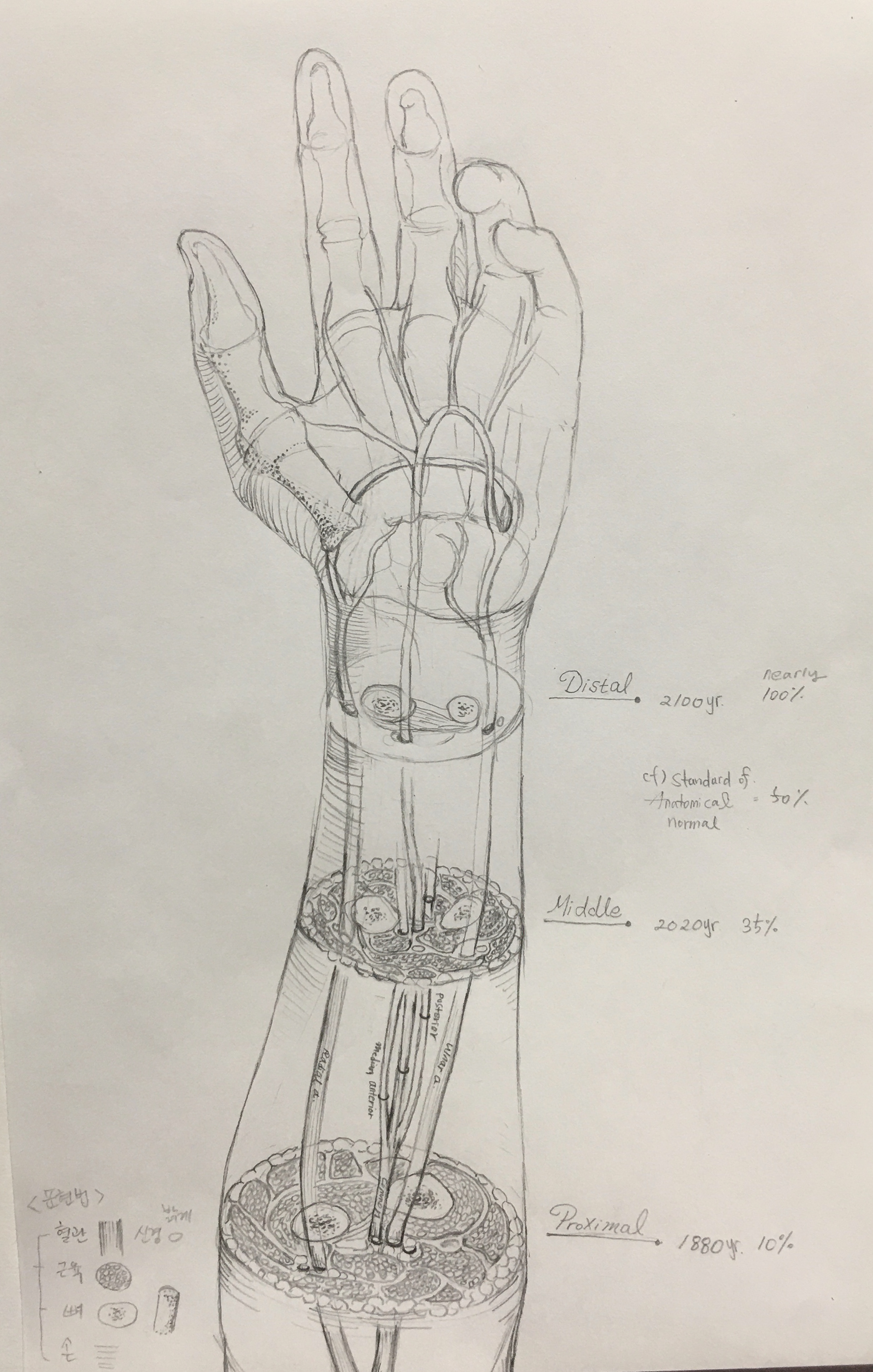
Median Artery_Sketch2
Median Artery_Sketch3
Median Artery_Sketch4